Department of Surgery, School of Medicine, Kyungpook National University, Taegu, Korea.
Correspondence: Jinyoung Park, M.D., Department of Surgery, Kyungpook National University Hospital, 50 Samduk-2 Ga, Chung-gu, Taegu 700-721, Korea. Tel: 053)420-5612, Fax: 053)421-0510, kpnugs@yahoo.co.kr
• Received: June 13, 2012 • Accepted: September 6, 2012
Although hemangiomas are common vascular tumors that can occurany where in the body, they seldom involve the gastrointestinal tract. Hemangiomas of the gastrointestinal tract in infants and children are rare benign vascular tumors that most commonly present with gastrointestinal bleeding. We describe here the case of 2-year-old boy with intestinal bleeding caused by a large jejunal cavernous hemangioma, which was treated by laparoscopy-assisted resection of the affected portion of the jejunum.
2. Levy AD, Abbott RM, Rohrmann CA Jr, Frazier AA, Kende A. Gastrointestinal hemangiomas: Imaging findings with pathologic correlation in pediatric and adult patients. AJR. 2001. 177:1073-1081.
3. Magnano A, Privitera A, Calogero G, Nanfito L, Basile G, Sanfilippo G. Solitary hemangioma of the small intestine: an unusual cause of bleeding diagnosed at capsule endoscopy. J Pediatr Surg. 2005. 40:E25-E27.
4. Jarvi K, Roebuck DJ, Sebire NJ, Lindley K, Shah N, Salomon J, Curry JI. Successful treatment of extensive infantile hemangiomatosis of the small bowel in a 3-month-old with thalidomide and somatostatin analog. J Pediatr Gastroenterol Nutr. 2008. 46:593-597.
5. Choi YH, Kim YH, Kim JW, Kim HY, Lee SH, Lee BY, Seo YS, Ham JH, Chung IK, Kim HS, Lee MH, Kim SJ, Cho HD. A Case of Giant Cavernous Hemangioma of Transverse Colon Associated with Hematochezia. Korean J Gastrointest Endosc. 2002. 25:484-488.
6. Chattopadhyay A, Kumar V, Maruliah M, Rao PL. Duodenojejunal obstruction by a hemangioma. Pediatr Surg Int. 2002. 18:501-502.
7. Chen CH, Jones J, McGowan P. Profound iron deficiency anemia caused by a small-intestinal cavernous hemangioma. Gastrointest Endosc. 2009. 69:1392-1393.
8. Lee YJ, Bae SH, Song ES, Choi SJ, Kim YH, Choi YY. A Case of Intestinal Hemangioma Complicated with Thrombocytopenia (Kasabach-Merritt syndrome) in Premature Infant. J Korean Soc Neonatol. 2010. 17:116-122.
9. Singh BP, Kumar A, Chattopadhyay TK. Intussuscepting ileal hemangioma with perforation. Indian J Gastroenterol. 1992. 11:94-95.
10. Jeong CY, Jung EJ. An Ileocolic Intussusception Caused by Small Bowel Hemangioma. J Korean Surg Soc. 2004. 67:490-492.
11. Sakaguchi M, Sue K, Etoh G, Takagishi T, Ezaki T, Nakamura M, Yamanaka K, Morita J. A case of solitary cavernous hemangioma of the small intestine with recurrent clinical anemic attacks in childhood. J Pediatr Gastroenterol Nutr. 1998. 27:342-343.
12. Kavin H, Berman J, Martin TL, Feldman A, Forsey-Koukol K. Successful wireless capsule endoscopy for a 2.5-year-old child: obscure gastrointestinal bleeding from mixed, juvenile, capillary hemangioma-angiomatosis of the jejunum. Pediatrics. 2006. 117:539-543.
13. Lee HS, Heo SY, Kim WD. Successful Management with Vincristine after Failure of Prednisolone Therapy for Diffuse Neonatal Hemangiomatosis. Korean J Pediatr. 2005. 48:1004-1008.
14. Ahn BY, Lee DH, Kim HC, Kang GH, Kim JC. Hormonal Treatment of Intestinal Cavernous Hemangioma Report of 2 cases. J Korean Soc Coloproctol. 2000. 16:34-36.
Fig. 1
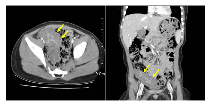
Abdominopelvic CT scan with contrast medium, showing circumferential thickening of the small bowel with central enhancement in the pelvic cavity.
Fig. 2
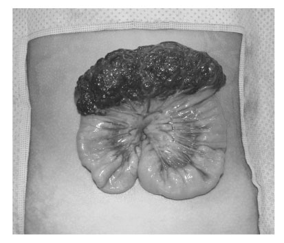
Surgical specimen, showing a 10 × 4 cm circumferential blood-filled reddish mass involving the serosal surface of the jejunum.
Fig. 3
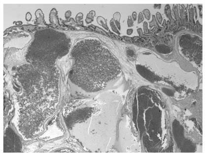
Microscopic examination of the hemangioma, showing large, dilated, blood-filled vessels lined by flattened endothelium and focal thickening of the vascular walls by adventitial fibrosis (H & E, × 20 original magnification).
Large Cavernous Hemangioma in the Jejunum of a 2-year-old Boy Treated by Laparoscopy-assisted Resection
Fig. 1
Abdominopelvic CT scan with contrast medium, showing circumferential thickening of the small bowel with central enhancement in the pelvic cavity.
Fig. 2
Surgical specimen, showing a 10 × 4 cm circumferential blood-filled reddish mass involving the serosal surface of the jejunum.
Fig. 3
Microscopic examination of the hemangioma, showing large, dilated, blood-filled vessels lined by flattened endothelium and focal thickening of the vascular walls by adventitial fibrosis (H & E, × 20 original magnification).
Fig. 1
Fig. 2
Fig. 3
Large Cavernous Hemangioma in the Jejunum of a 2-year-old Boy Treated by Laparoscopy-assisted Resection