Purpose Crohn’s disease (CD) is mainly presenting gastrointestinal symptoms but also may involve perianal diseases, with anal fistula being the most common. Anal fistula surgery performed without recognition that the patient has CD increases the complications such as delayed wound healing and anal sphincter injury. This study aimed to investigate clinical symptoms and surgical findings that could allow early diagnosis of CD in patients aged 10 to 19 years who underwent anal fistula surgery.
Methods Among the 320 patients under the age of 19 who underwent anal fistula surgery, those who were diagnosed with tuberculosis fistula were excluded, medical records of 316 patients were examined. We investigated the characteristics of anal fistula and postoperative wounds as well as the colonoscopic and surgical findings.
Results Compared to 272 patients not diagnosed with CD (non-CD group), 44 patients diagnosed with CD (CD group) showed significantly higher levels of C-reactive protein as well as anal discharge, family history of inflammatory bowel disease, history of anal fistula, diarrhea, abdominal pain, weight loss, complex anal fistula, recurred anal fistula, delayed wound healing and friable/edematous/granulation of surgical site.
Conclusion For patients aged 10–19 years with anal fistulas, emphasizing early suspicion and an active diagnostic work-up is essential for early diagnosis of CD.
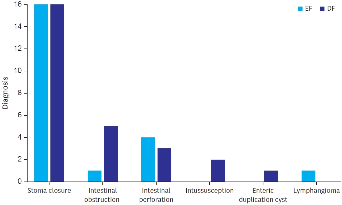
Purpose The establishment of enteral feeding is the end point of any intestinal anastomosis. This study examined the effects of early feeding (EF) as compared to delayed feeding (DF) on postoperative outcomes after intestinal anastomosis in children.
Methods This was a randomized controlled pilot study to assess the effect of EF vs. DF in terms of time to reach full feed, along with wound infection and anastomotic leak.
Results Twenty-eight patients were enrolled in both study groups. The median time to first feed in EF was 60 hours and 96 hours in DF. The median time to first bowel sound was 42 hours in EF and 48 hours in DF (p=0.208). The median time to first bowel movement was 72 hours in EF and 72 in DF (p=0.820). The median time of postoperative hospital stay was 5.5 days in EF and 6.0 days in DF (p=0.01). There was no significant difference in complications of wound infection, wound dehiscence, relook surgery, or anastomotic leak in both groups.
Conclusion EF after intestinal anastomosis is safe and feasible in children after intestinal anastomosis.
Citations
Citations to this article as recorded by
Modified Enhanced Recovery After Surgery Protocols in Pediatric Gastric Transposition: Effects on Recovery and Outcomes Mohit B. Chauhan, Nitin James Peters, Muneer Abas Malik, Shivani Dogra, Ravi Prakash Kanojia, Rajni Sharma, Sandhya Yaddanapudi, Monika Bawa, Shailesh Solanki, Jai Kumar Mahajan Journal of Indian Association of Pediatric Surgeons.2026; 31(1): 20. CrossRef
Early vs. delayed feeding after pediatric gastrointestinal surgery: a systematic review and meta-analysis Mohammed Al Blooshi Pediatric Surgery International.2026;[Epub] CrossRef
Nutritional Timing in Pediatric Stoma Reversal: Early Versus Late Feeding Practices Nadia Shoukat, Mumtaz Ahmed Qureshi, Ali Hasham, Sadia Shoukat, Nazia Azam Yousfani, . Matiullah Biological and Clinical Sciences Research Journal.2025; 6(9): 85. CrossRef
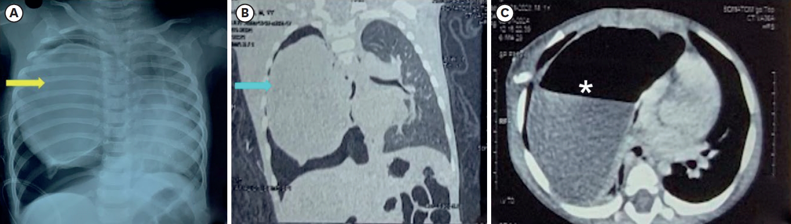
Intrapulmonary bronchogenic cysts are rare congenital anomalies that often present diagnostic and management challenges due to nonspecific symptoms. We report a one-year-old female with progressive respiratory distress who was initially misdiagnosed with pneumothorax. Imaging revealed a large intrapulmonary cyst, and surgical excision confirmed the diagnosis. Early intervention ensured a favorable outcome, with no recurrence in one year. This case highlights the importance of considering bronchogenic cysts in pediatric respiratory distress and emphasizes the value of timely surgical management.
Purpose The International Society for the Study of Vascular Anomalies (ISSVA) classification is crucial in diagnosing vascular anomalies (VAs), surpassing the International Classification of Diseases 10th Revision. This study aims to reevaluate diagnoses using ISSVA criteria and explore diagnostic patterns.
Methods Analyzing 138 pediatric VA patients diagnosed via magnetic resonance imaging from 2018 to 2023 at Asan Medical Center, we reviewed clinical, imaging, pathology, and genetic data. Diagnoses were revised per 2018 ISSVA criteria, assessing discrepancies.
Results Among 133 VA cases, 125 were malformations and eight were tumors, mostly in the head and neck. Clinical and imaging diagnoses disagreed in 51 cases. Some initially complex malformations were simplified. Lymphatic malformation cases shifted to VAs and vascular tumors were identified post-initial diagnosis.
Conclusion Accurate diagnosis of VAs is essential for prognosis, treatment planning, and predicting outcomes. However, 14.2% of patients showed discordance between clinical diagnoses and imaging findings. Capillary malformations were often overlooked in imaging but became evident with relevant clinical findings. Adopting a multidisciplinary approach and a unified diagnosis based on ISSVA classification is crucial for clearly defining VAs.
Purpose Complicated vascular anomalies, characterized by encasing vital organ or diffusely locating unresectable lesion, pose therapeutic challenges with limited response to conventional treatment such as surgical resection or sclerotherapy. Sirolimus, an mammalian target of rapamycin inhibitor, has shown promising therapeutic effects in patients with vascular anomalies by inhibiting vascular endothelial growth factor, as reported in several studies. Here, we analyzed the treatment outcomes of patients who received sirolimus for complicated vascular anomalies at our institution.
Methods Patients treated with sirolimus at the Department of Pediatric Surgery, Asan Medical Center from January 2018 to December 2021 were included. Sirolimus was administered twice daily at a dose of 0.8 mg per body surface area (BSA), with dose adjustments to achieve a target drug concentration of 8–12 ng/mL. Adverse drug effects and therapeutic responses were periodically assessed. Treatment efficacy was evaluated based on clinical findings pre- and post-sirolimus administration, absolute volume reduction of lesions through imaging tests (magnetic resonance imaging; MRI), and relative volume reduction adjusted to the patient's BSA.
Results There were 16 females (50.0%) and 16 males (50.0%), with a median follow-up period of 41 months after sirolimus administration. Vascular anomaly types included lymphatic malformations (41%), venous malformations (28%), lymphovenous malformations (19%), and others (12.5%). The most common adverse effect was oral ulcer (6 patients). MRI volumetry revealed volume decreases in 17 patients (53.1%) with 22 patients (71%) exhibited lesion decreases relative to BSA. Notably, 9 patients (28.1%) had markedly decreased volume reduction based on absolute volume, and 12 (38.7%) based on volume compared to BSA.
Conclusion Over a 2-year follow-up, sirolimus was effective in treating patients with complicated vascular anomalies, when administered with cautious consideration of side effects. A multidisciplinary approach is needed for evaluating treatment outcomes in these patients, necessitating further long-term research on adverse effects.
Purpose Most patients with perinatally detected subhepatic cysts receive information suggestive of a suspected diagnosis of choledochal cyst (CC). However, it is not uncommon to be finally diagnosed with cystic biliary atresia (CBA), a disease with a more unfavorable prognosis. This study aimed to investigate the distribution of the final diagnoses of perinatally detected subhepatic cysts and to compare patients diagnosed with CC and CBA.
Method We performed a retrospective review of patients with subhepatic cysts detected using ultrasonography during perinatal period, between January 2012 and December 2022.
Result This retrospective study included 52 patients with perinatal subhepatic cysts. Of these, 71.2% (37/52) were diagnosed with CC, 15.4% (8/52) with CBA, and 5.8% (3/52) with duplication of the alimentary tract. Only 1.9% (1/52) of the patients were diagnosed with biliary atresia, gallbladder duplication, mesenteric lymphatic malformation, or were normal. Of all patients, 86.5% (45/52) were diagnosed with CC or CBA, with CBA accounting for 17.8% (8/45). There were no statistically significant differences between the CC and CBA groups regarding the gestational age at which the cyst was first detected and the final size of the cyst measured on prenatal ultrasound.
Conclusion Subhepatic cysts detected during the perinatal period are typically diagnosed as CC. However, this study revealed that 15.4% of all patients were diagnosed with CBA, despite no significant differences in prenatal ultrasound findings. Therefore, it is essential to consider the possibility of CBA in cases of perinatally detected subhepatic cysts.
Citations
Citations to this article as recorded by
Prenatal Diagnosis of Isolated Caroli Disease Caused by a Homozygous PKHD1 Variant: A Case Report and Literature Review Hai Wang, Zitong Xu, Xianjue Zheng, Haojie Pan, Yimin Wang, Haiying Chen, Zhenzhen Zheng, Hongping Zhang, Jiayong Zheng Clinical and Experimental Obstetrics & Gynecology.2026;[Epub] CrossRef
Jinyoung Park, Dayoung Ko, Eun-jung Koo, Hyunhee Kwon, Ki Hoon Kim, Dae Yeon Kim, Seong Chul Kim, Soo-Hong Kim, Wontae Kim, HaeYoung Kim, Hyun-Young Kim, So Hyun Nam, Jung-Man Namgoong, Junbeom Park, Taejin Park, Min-Jung Bang, Jeong-Meen Seo, Ji-Young Sul, Joonhyuk Son, Joohyun Sim, Soo Min Ahn, Hee-Beom Yang, Jung-Tak Oh, Chaeyoun Oh, Joong Kee Youn, Sanghoon Lee, Ju Yeon Lee, Kyong Ihn, Hye Kyung Chang, Yeon Jun Jeong, Eunyoung Jung, Jae Hee Chung, Min Jeong Cho, Yun-Mee Choe, Seok Joo Han, In Geol Ho, Jeong Hong
Adv Pediatr Surg 2025;31(1):8-15. Published online May 28, 2025
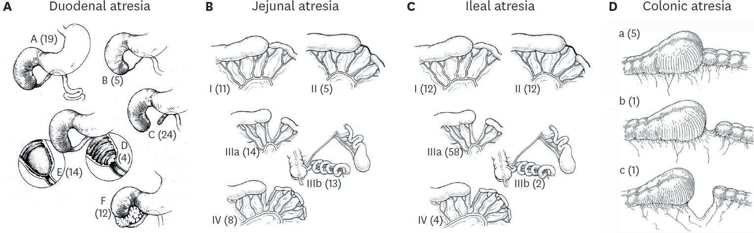
Purpose This study aims to investigate and compare the incidence, demographic characteristics, clinical manifestations, preoperative diagnostic methods, anatomical classifications, associated anomalies, operative treatments, and postoperative outcomes of patients with intestinal atresia treated by the members of the Korean Association of Pediatric Surgeons (KAPS) through three nationwide surveys.
Methods KAPS conducted 3 national surveys in 1998, 2010, and 2024 to examine the patients diagnosed with intestinal atresia. In preparation for the survey, we developed a customized case registration form to obtain data on patient sex, birth weight, gestational age, clinical manifestations, preoperative diagnostic methods, anatomical types, associated anomalies, operative treatments, and postoperative outcomes. Authorized KAPS members completed the case registration form.
Results The first, second, and third national surveys included 218, 222, and 236 individuals diagnosed with intestinal atresia, respectively. The male-to-female ratios were 1.5:1, 1.1:1, and 1.1:1, respectively. The first, second, and third national surveys revealed that 34.3%, 43.3%, and 53.4% of patients were born before 37 weeks of gestation, respectively. Additionally, 28.7%, 32.0%, and 40.7% of patients had a birth weight under 2,500 g. In the third national survey, duodenoduodenostomy was the most common procedure, performed in 70 out of 82 patients diagnosed with duodenal atresia. Resection and anastomosis were the main surgical procedures conducted in 47 out of 54 cases of jejunal atresia and 74 out of 92 cases of ileal atresia. The mortality rates in the first, second, and third national surveys were 13.8%, 3.6%, and 1.3% respectively, with the lowest rate observed in the third national survey.
Conclusion These national surveys offer valuable insights into the current state of intestinal atresia, including specific surgical interventions and postoperative outcomes in South Korea. For pediatric surgeons aiming to enhance their understanding of intestinal atresia and its treatment options, these surveys could be an indispensable resource and guide.
A ciliated foregut cyst is a rare developmental anomaly. It develops from the primitive foregut. It is usually located supra-diaphragmatically. Its localization in the gallbladder is very infrequent and has been sparsely reported. We report a rare case of a ciliated cyst of the gallbladder in an 11-year-old female, who presented with complaints of upper abdominal pain for 2 months. She was suspected to have gallbladder duplication or gallbladder diverticulum on imaging. The histopathology reported this anomaly as a ciliated foregut cyst. The ciliated cyst of the gallbladder is a benign congenital lesion. Abdominal ultrasonogram and computed tomography/magnetic resonance imaging are suggestive of a cystic lesion of the gallbladder. The definitive diagnosis is by histopathological examination. This is a rare clinicopathological condition in the pediatric age group. The recommended treatment is laparoscopic cholecystectomy. The role of conservative management has not been established due to the rarity of the condition.
Yeon Jun Jeong, Dayoung Ko, Hyunhee Kwon, Ki Hoon Kim, Dae Yeon Kim, Soo-Hong Kim, Wontae Kim, Hae-Young Kim, Hyun Young Kim, Seong Chul Kim, Younghyun Na, Jung-Man Namgoong, So Hyun Nam, Junbeom Park, Jinyoung Park, Tae-Jun Park, Jeong-Meen Seo, Ji-Young Sul, Joonhyuk Son, Hyun Beak Shin, Joohyun Sim, Soo Min Ahn, Hee Beom Yang, Jung-Tak Oh, Chaeyoun Oh, Joong Kee Youn, Sanghoon Lee, Ju Yeon Lee, Kyong Ihn, Hye Kyung Chang, Eunyoung Jung, Jae Hee Chung, Yu Jeong Cho, Yun Mee Choe, Soo Jin Na Choi, Seok Joo Han, In Geol Ho, Ji-Won Han
Adv Pediatr Surg 2025;31(2):47-58. Published online July 16, 2025
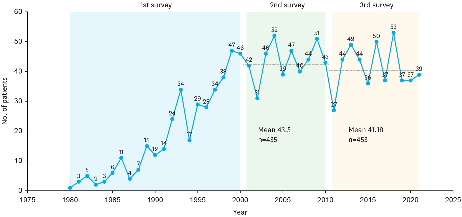
Purpose Biliary atresia (BA) is a rare but progressive cholangiopathy and the leading cause of pediatric liver transplantation worldwide. The Korean Association of Pediatric Surgeons (KAPS) has conducted three national surveys (2001, 2011, and 2023) to assess long-term trends in the diagnosis, treatment, and outcomes of BA. This study provides a comparative analysis of the 2nd and 3rd national surveys, with reference to selected findings from the 1st survey.
Methods This study included 453 patients from the 3rd national survey (2011–2021) and 435 patients from the 2nd survey (2001–2010), all of whom underwent Kasai portoenterostomy. Data were collected via electronic case report forms from pediatric surgical centers nationwide. Comparisons were made regarding demographics, clinical features, diagnostic patterns, operative details, follow-up outcomes, and survival. Kaplan–Meier analysis was used to evaluate long-term survival.
Results The mean number of BA patients per year remained stable between surveys (43.5 in the 2nd, 41.18 in the 3rd), though centralization of care increased, with 61.5% of cases managed by two major institutions in the 3rd survey. The median age at surgery decreased, and the use of preoperative imaging (especially magnetic resonance cholangiopancreatography) increased. The 10-year native liver survival rate declined from 59.8% to 53.7%, while overall 10-year survival improved slightly (92.9% to 93.2%). Postoperative complications, such as cholangitis and liver failure, persisted but were better categorized. The 3rd survey also reported improved mortality (4.9%) and reduced follow-up loss (11.5%) compared to the 2nd survey.
Conclusion While overall survival after Kasai operation has remained high and even improved, native liver survival has slightly declined. The findings reflect earlier diagnosis, more consistent diagnostic imaging, and increasing centralization of care. These trends underscore the importance of long-term nationwide data collection in guiding future strategies for BA management in Korea.
Hepatobiliary ascariasis (HA) is a rare condition associated with significant morbidity. Laparoscopic extraction of Ascaris lumbricoides from the biliary tract is a safe approach in patients who do not improve with antihelminthic treatment and when retrograde endoscopic cholangiography is not feasible or when not all nematodes can be removed using this method. Here I present the technique used in two pediatric patients with HA.
Hee-Beom Yang, Soo Min Ahn, Min Jeng Cho, Yong-Hoon Cho, Soo Jin Na Choi, Yoon Mi Choi, Jae Hee Chung, Seok Joo Han, In Geol Ho, Jeong Hong, Kyong Ihn, Yeon Jun Jeong, Eunyoung Jung, Dae Youn Kim, Hae-Young Kim, Ki Hoon Kim, Seong Chul Kim, Soo-Hong Kim, Eun-Jung Koo, Hyun Hee Kwon, Yong Jae Kwon, Nam-Hyuk Lee, Ju Yeon Lee, Sanghoon Lee, Jung-Man Namgoong, Chaeyoun Oh, Jung-Tak Oh, Jin Young Park, Junbeom Park, Jeong-Meen Seo, Jae Ho Shin, Hyun Beak Shin, Joohyun Sim, Jiyoung Sul, Joon Kee Youn, Hyun-Young Kim
Adv Pediatr Surg 2025;31(2):66-76. Published online November 25, 2025
Purpose To report a nationwide survey on neuroblastoma conducted by the Korean Association of Pediatric Surgeons (KAPS) in 2020.
Methods The clinical data of pediatric patients diagnosed with and treated for neuroblastoma from 2005 to 2019 in 19 hospitals of KAPS members were collected. Survival and prognostic factor analyses were performed using the log rank test and Cox proportional hazard analysis. A p-value <0.05 was considered significant.
Results A total of 669 patients with neuroblastoma were registered for the study. The results were presented and discussed at the 36th annual meeting of the KAPS, which was held in Seoul on August 21, 2020.
Conclusion This study provides information on patient demographics, prognostic outcomes, and comprehensive treatment outcomes for neuroblastoma. The study is expected to be an important reference for improving pediatric surgeons’ understanding and treatment of neuroblastoma.
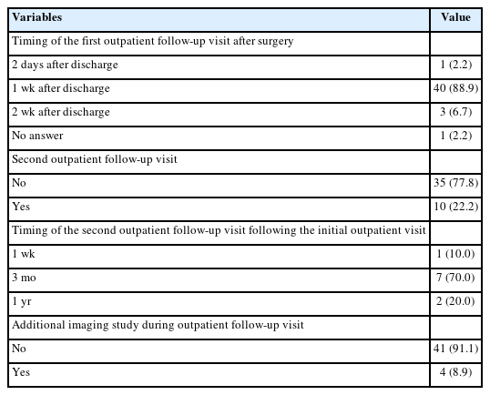
Purpose This study aimed to evaluate postoperative outpatient follow-up practices among pediatric surgeons in Korea for five common congenital diseases: esophageal atresia with tracheoesophageal fistula (EA/TEF), anorectal malformation (ARM), Hirschsprung’s disease (HSCR), choledochal cyst (CC), and inguinal hernia (IH).
Methods A web-based survey consisting of 43 questions was distributed to members of the Korean Association of Pediatric Surgeons. The survey assessed the timing, frequency, and duration of outpatient follow-up, as well as disease-specific practices.
Results Of 154 invited surgeons, 45 (29.2%) responded. Most scheduled the first follow-up visit within one week after discharge. During the first postoperative year, follow-up visits were commonly held every three months, followed by six months or annual intervals. Most surgeons concluded follow-up before age 18; however, 15.6%–37.8% reported continuing follow-up into adulthood depending on the disease. Variation was observed in disease-specific practices: 44.4% routinely performed contrast studies for EA/TEF follow-up; sizes #14–15 Hegar dilators were most used in ARM; only 6.7% performed routine rectal irrigation in HSCR. For CC, 88.9% checked both blood tests and ultrasonography. Most IH patients received only one follow-up visit.
Conclusion While early postoperative follow-up practices among pediatric surgeons in Korea appear relatively consistent, wide variation exists in long-term strategies and disease-specific protocols. This reflects the tendency to rely on individual clinical judgment and highlights the need for standardized, national consensus.
Citations
Citations to this article as recorded by
Transition of Care to Late Adolescent/Adults in Paediatric Surgery: Perspectives From Indian Paediatric Surgeons Sunita Singh, Yogesh Kumar Sarin, Subhashis Saha, Mukesh Shukla Journal of Indian Association of Pediatric Surgeons.2026; 31(4): 541. CrossRef
Intestinal failure (IF) is a term used to define the state where intestine’s function is significantly reduced, to the point where adequate growth and hydration cannot be maintained. In such cases, intravenous nutritional support is essential for sustaining the patient’s life. In pediatric patients, the most common cause of IF is short bowel syndrome (SBS). Due to the prolonged treatment and high complication rates, management of SBS remains a continuous challenge to many physicians. Herein, we report the case of a 2,260 g premature female infant born at 35-week gestational age with type 4 jejunoileal atresia. She presented with ultrashort bowel syndrome, having a bowel length of less than 15 cm, but ultimately achieved gut autonomy and restored bowel function through successful intestinal rehabilitation within the first two years of life.
Yeon Jun Jeong, Dayoung Ko, Eun-Jung Koo, Hyunhee Kwon, Dae Yeon Kim, Soo-Hong Kim, Wontae Kim, Hae-Young Kim, Hyun Young Kim, Seong Chul Kim, Younghyun Na, Jung-Man Namgoong, So Hyun Nam, Sungjoo Park, Junbeom Park, Jinyoung Park, Tae-Jun Park, Jeong-Meen Seo, Ji-Young Sul, Joonhyuk Son, Hyun Beak Shin, Joohyun Sim, Jung-Tak Oh, Chaeyoun Oh, Joong Kee Youn, Sanghoon Lee, Ju Yeon Lee, Cheolgu Lee, Kyong Ihn, Eunyoung Jung, Jae Hee Chung, Yong-Hoon Cho, Yun Mee Choe, Soo Jin Na Choi, Seok Joo Han, In Geol Ho
Adv Pediatr Surg 2024;30(2):39-51. Published online December 13, 2024
Purpose This study provides insights into the prevalence at birth, clinical characteristics, and outcomes of gastroschisis and omphalocele in Korea over the past decade, addressing the lack of localized data despite advanced healthcare capabilities.
Methods The study retrospectively analyzed data from 20 pediatric surgical centers in Korea from January 2012 to December 2021, including 269 patients diagnosed with gastroschisis or omphalocele. Data variables included gender, gestational age, birth weight, associated anomalies, type of defect, surgical interventions, and outcomes.
Results The study covered 269 patients, with 80 gastroschisis and 189 omphalocele cases. Gastroschisis prevalence at birth remained stable at 2.15 per 100,000 live births, while omphalocele increased to 5.08 per 100,000. Both conditions had similar gender ratios (0.95). Gastroschisis patients had lower birth weights (2,463.90±505.50 g) and smaller head circumferences (31.97±1.86 cm) compared to omphalocele patients (2,757.65±761.24 g, 32.78±2.64 cm). Omphalocele cases had more associated anomalies, especially cardiovascular issues. Prenatal diagnosis rates were high: 93.7% for gastroschisis and 86.4% for omphalocele. About 96.3% of gastroschisis and 84.1% of omphalocele patients were born in their treatment hospitals. Gastroschisis patients underwent surgery sooner (average 3.5 days) and started feeding later (16.5 days) than omphalocele patients (average 56.5 days to surgery, 6.6 days to start feeding). Hospital stays and follow-up durations were similar, averaging around 782.6 days for gastroschisis and 800.3 days for omphalocele patients. Survival rates were 89.7% for gastroschisis and 87.1% for omphalocele.
Conclusion The study highlights the need for early diagnosis, centralized care, and specialized surgical approaches to optimize outcomes for gastroschisis and omphalocele patients in Korea. Enhanced prenatal screening and surgical protocols are recommended to improve these patients' prognosis.
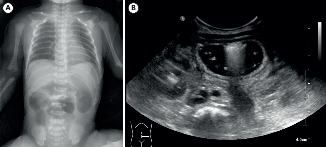
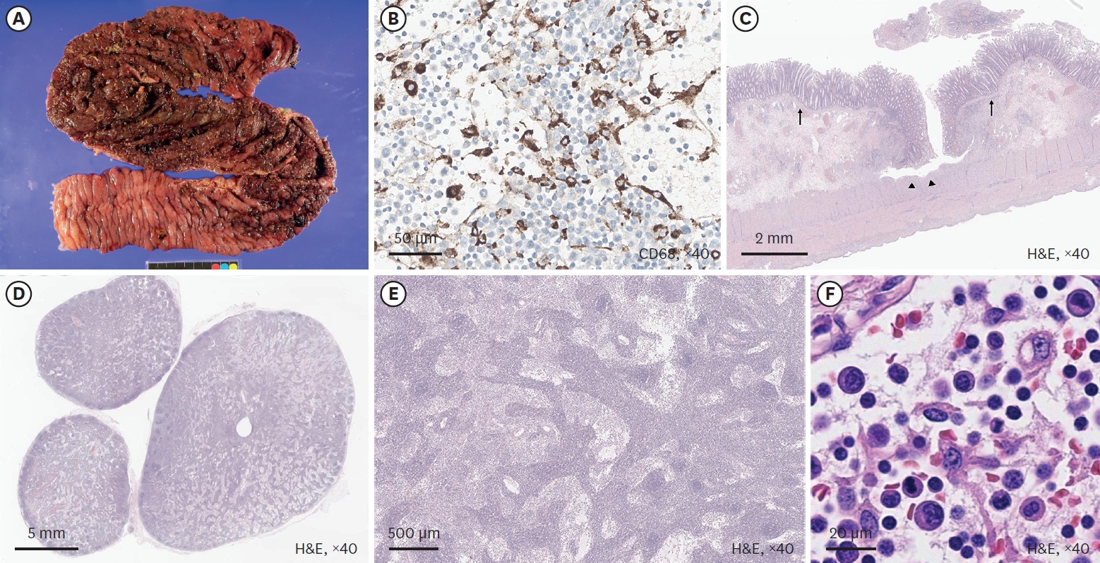
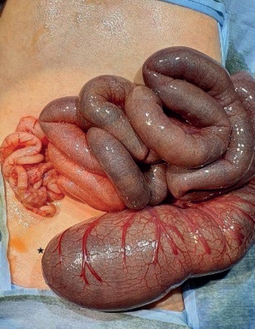
Pediatric intestinal perforation is a surgical emergency that must be promptly addressed regardless of the specific cause. Here we report a case of colon perforation caused by indeterminate inflammatory bowel disease (IBD) in an autistic 13-year-old boy. Ulcerative colitis (UC) and lymphoma were first suspected but subsequently ruled out. The patient was previously hospitalized locally for 8 days due to diarrhea. He was diagnosed with UC and colon perforation in the emergency room. He then underwent subtotal colectomy with end ileostomy. Pathological examination of the colon showed multiple perforations with absence of chronic crypt change (a characteristic of UC), presence of undermining ulcers, and atypical lymphocyte infiltrations. Lymphoma was ruled out from immunohistochemistry and blood tests. Indeterminate colitis was finally suggested as the cause of perforation. Genetic analysis confirmed KBG syndrome, but no abnormalities otherwise known to be relevant to colitis. This case demonstrates that spontaneous colon perforation might occur in KBG syndrome patients suffering from severe enteritis without IBD, malignancy, or other conditions known to cause perforation, supporting the necessity of close monitoring when such patients present with severe symptoms including fever and abdominal distension without showing improvement.
Proliferative myositis (PM) is a rare benign soft tissue neoplasm with a distinctive pseudosarcomatous proliferative reaction of muscles in tumors. Its rapid growth and bizarre microscopic appearance often require a differential diagnosis from a sarcomatous lesion. It has been reported occasionally, mostly as case reports in adult patients. Herein, we present a neonatal case of PM. To the best of our knowledge, this is the first report in the neonatal period.
The concurrent occurrence of colonic atresia, malrotation, and Hirschsprung’s disease in neonates is extremely rare. These anomalies often share embryologic origins and present overlapping clinical symptoms that complicate diagnosis and management. We report two neonatal cases with this rare triad. Case 1 involved a term neonate initially diagnosed with esophageal atresia and later found to have colonic atresia, malrotation, and Hirschsprung’s disease. Case 2 was a preterm neonate presenting with abdominal distension and perforation, ultimately diagnosed with the same triad. Both underwent staged surgical management, including Duhamel’s procedures after confirming aganglionosis. Awareness of the possible coexistence of these anomalies is essential in neonates with colonic atresia and non-fixed colon. Surgical planning should anticipate aganglionosis and include rectal biopsy. This report emphasizes the importance of early suspicion and multidisciplinary approach for optimal outcomes.
Hirschsprung disease (HSCR) is a genetic disorder with an incidence of 1:5000, seen in the pediatric age group. The association between HSCR and neuroblastoma (NBL), ends of the neurocristopathy spectrum is rare. Less than 10 cases of this association are reported in the literature and the association between the Phox gene and Sox10 gene in the pathophysiology of these is being studied. We report a one-year-old baby, who presented to us, with chronic constipation on regular enemas and laxative usage. There was a history of delayed passage of meconium. At the time of Duhamel’s pull through a well-defined, bilobed hard presacral mass, was encountered. Excision and coccygectomy were done and the pull was completed. The histopathology showed a well-differentiated NBL. Fludeoxyglucose positron emission tomography scan and the N-Myc amplification were negative and the patient was managed with expectant treatment. She is doing well over a 3-year follow-up with no recurrence and good resolution of bowel functions.
Since the first introduction of robotic surgery systems in Korea in 2005, there has been a gradual increase in the number of robotic surgeries performed. However, robotic liver resection is one of the most complex procedures, and its application, especially to children, is still limited. Therefore, in this study, we aim to present our experiences with 2 pediatric patients who underwent robotic liver resection in Asan Medical Center and discuss the safety and feasibility of robot-assisted hepatectomy in pediatrics.
Hee-Beom Yang, Min Jeng Cho, Yu Jeong Cho, Yoon Mi Choi, Jae Hee Chung, Seok Joo Han, Jeong Hong, Eunyoung Jung, Ki Hoon Kim, Soo-Hong Kim, Cheol-Gu Lee, Nam-Hyuk Lee, Ju Yeon Lee, Sanghoon Lee, Suk Bae Moon, Young-Hyun Na, So Hyun Nam, Chaeyoun Oh, Jin Young Park, Junbeom Park, Tae-Jin Park, Jae Ho Shin, Joonhyuk Son, Hyun-Young Kim, The Korean Association of Pediatric Surgeons
Adv Pediatr Surg 2025;31(2):59-65. Published online August 5, 2025
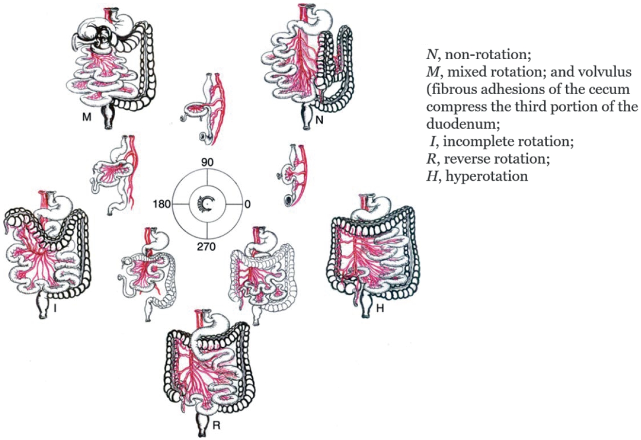
Purpose To report the findings of a perception survey on intestinal malrotation conducted by the Korean Association of Pediatric Surgeons (KAPS) in 2021.
Methods The perceptions on intestinal malrotation regarding clinical decision making of the KAPS members were collected through web-based survey.
Results A total of 22 surgeons were answered for this study. The results were presented and discussed at the 37th annual meeting of KAPS, which was held in Seoul on June 18, 2021.
Conclusion This study provides the clinical decisions of the KAPS members on the intestinal malrotation. The study is expected to be an important reference for improving pediatric surgeons’ understanding and treatment of intestinal malrotation.
Duodenal web (DW) is a rare congenital anomaly of the intestinal tract that can lead to severe dehydration and electrolyte imbalance. A 14-day-old boy presented with recurrent vomiting and weight loss and was diagnosed with DW. Duodenoscopy identified a pinhole structure in the second portion of the duodenum, prompting a subsequent endoscopic balloon dilatation procedure. Following the procedure, vomiting and abdominal distension resolved, and the patient was discharged on a regular diet. No symptoms recurred during follow-up.
Purpose Enteric duplication cysts (EDCs) are rare congenital anomalies of the gastrointestinal tract. This study aimed to delineate the clinical, anatomical, and pathological spectrum of EDCs based on a 40-year single-center experience.
Methods A retrospective review was conducted of 45 pediatric patients who underwent surgical treatment for EDCs at a single institution between 1985 and 2023. Clinical records, imaging studies, and pathological reports were analyzed.
Results The study included 28 males and 17 females, with a median age at surgery of 4.7 months. Most patients (75.6%) underwent surgery before 2 years of age. The ileum was the most common location (57.8%), followed by the jejunum (11.1%) and ileocecal valve (11.1%). Vomiting (46.7%) was the most common presenting symptom. Emergency surgery was required in 28.9% of cases because of complications such as volvulus or intussusception. Preoperative imaging using ultrasonography (US) and/or computed tomography resulted in a correct diagnosis in 34 of 45 patients (75.6%), with EDCs correctly identified in 30 patients (66.7%), frequently based on the characteristic “double wall sign” observed on US. Histopathological examination identified heterotopic gastric mucosa in 61.4% of evaluable cases. Postoperative outcomes were generally favorable, with a median hospital stay of 7.5 days.
Conclusion EDCs are rare congenital anomalies that are primarily diagnosed during early childhood. The ileum is the most frequent site of involvement, and clinical presentation is often related to acute complications. Prompt and complete surgical excision remains the definitive treatment and leads to favorable short-term postoperative outcomes, with no recurrence observed during the available follow-up period.
Jinyoung Park, Dae Yeon Kim, Seong Chul Kim, Hyun-Young Kim, So Hyun Nam, Jeong-Meen Seo, Jung-Tak Oh, Myung-Duk Lee, Suk-Koo Lee, Soo Min Ahn, Hye Kyung Chang, Sung Eun Jung, Yeon Jun Jeong, Eunyoung Jung, Jae Hee Chung, Yong Hoon Cho, Soon Ok Choi, Seung Hoon Choi, Yun Mee Choe, Seok Joo Han, Jeong Hong, Nam-Hyuk Lee
Adv Pediatr Surg 2024;30(1):1-8. Published online May 31, 2024
Purpose The Korean Association of Pediatric Surgeons (KAPS) conducts annual nationwide surveys on various aspects of pediatric surgical diseases, with the results being discussed during KAPS’s annual spring meetings.
Methods KAPS conducted two national surveys, in 1995 and 2016, to investigate esophageal atresia (EA) with or without tracheoesophageal fistula (TEF). The authors analyzed data from these surveys to identify differences or changes in the annual occurrence, demographic characteristics, clinical presentation, preoperative diagnostic methods, anatomical type, associated anomalies, surgical treatment, and postoperative outcomes among patients with EA/TEF treated by KAPS members.
Results The first and second national surveys included 148 and 211 patients with EA/TEF, respectively. Excessive salivation was the most prevalent clinical symptom in both surveys. Type C was the most common form of EA/TEF in both surveys. The first survey included 126 patients, all of whom underwent open surgery. In the second survey, 152 (78.4%) of 194 patients underwent open surgery, while 34 (17.5%) underwent thoracoscopic surgery. Primary esophageal repair was performed on 96 (76.2%) of 126 patients in the first survey and on 160 (82.5%) of 194 patients in the second survey. Anastomotic strictures developed in 21.4% and 32.5% of patients, anastomotic leakage in 22.2% and 10.3%, recurrent fistula in 2.4% and 4.2% during the first and second surveys, respectively. The respective survival rates for group A were 90.2% and 98.3% in the first and second surveys. For group B, the rates were 73.9% and 98.1%, and for group C, they were 34.5% and 68.1%, respectively, according to the Waterston classifications.
Conclusion These nationwide surveys provide comprehensive information on the status, detailed treatment, and outcomes for Korean pediatric patients with EA/TEF. They are anticipated to be an invaluable resource and guide for pediatric surgeons seeking to expand their knowledge on EA/TEF and its treatment options.
This review summarizes the epidemiology, diagnosis, and management of pediatric foreign body (FB) ingestion, with particular emphasis on circumstances that may require surgical involvement. Initial evaluation includes history taking, assessment of symptoms, and radiographic imaging to determine the type and location of the ingested object. Plain radiography remains the primary diagnostic modality for detecting radiopaque objects and localizing them within the gastrointestinal tract; however, additional imaging may be needed for radiolucent objects or when complications are suspected. Management depends on the anatomical location and characteristics of the FB. Esophageal FBs generally require urgent endoscopic removal, especially when button batteries, magnets, or sharp objects are involved. After an object has passed into the stomach, many cases can be managed conservatively; however, high-risk objects, including button batteries, multiple magnets, and long or sharp items, may require early removal. FBs beyond the pylorus usually pass spontaneously but require monitoring for complications. Surgical intervention may be necessary when endoscopic removal is not feasible or when complications such as obstruction, perforation, or fistula formation occur. This review summarizes location- and object-specific management strategies and identifies situations in which early surgical involvement may improve outcomes in children with FB ingestion.
Morgagni hernia (MH) is a type of congenital diaphragmatic hernia that is rare and without any distinctive presentation. Chest radiographs can miss the diagnosis when solid organs instead of bowel loops are herniated. Echocardiography can perplex the diagnosis instead of aiding if MH is not suspected. We are here discussing the presentation and management of a neonate with MH, which was referred to our institute as a congenital heart disease.
Jinyoung Park, Dayoung Ko, Hyunhee Kwon, Dae Yeon Kim, Seong Chul Kim, Soo-Hong Kim, Wontae Kim, Hyun-Young Kim, So Hyun Nam, Jung-Man Namgoong, Sungjoo Park, Junbeom Park, Min-Jung Bang, Jeong-Meen Seo, Ji-Young Sul, Joohyun Sim, Soo Min Ahn, Hee-Beom Yang, Jung-Tak Oh, Chaeyoun Oh, Joong Kee Youn, Sanghoon Lee, Ju Yeon Lee, Cheolgu Lee, Kyong Ihn, Soo-Min Jung, Yeon Jun Jeong, Eunyoung Jung, Jae Hee Chung, Min Jeng Cho, Suhyeon Ha, Seok Joo Han, In Geol Ho
Adv Pediatr Surg 2026;32(1):18-26. Published online June 22, 2026
Purpose This study investigated the clinical characteristics, anatomical distribution, operative management, and postoperative outcomes of pediatric patients who underwent surgery for intestinal duplication and were registered through a nationwide multicenter survey conducted by the Korean Association of Pediatric Surgeons (KAPS).
Methods KAPS conducted a nationwide multicenter retrospective survey across 18 institutions between 2020 and 2024 and collected data from 144 patients.
Results Female patients accounted for 55.6% of surgically treated cases, corresponding to a male to female ratio of 1:1.25. Vomiting and abdominal pain were the most common presenting symptoms. Prenatal diagnosis was achieved in 43.7% of cases. The ileum was the most common site of intestinal duplication (41.0%). Cystic duplications predominated (82.6%), and communication with the native bowel was documented in 19.4% of cases. Elective surgery was performed in 83.3% of patients, with laparoscopic-assisted surgery being the most commonly used approach (52.8%). The most frequently performed surgical procedures were excision (49.3%) and bowel resection with anastomosis (47.2%). Recurrence occurred in three patients (2.1%), and mortality was reported in one patient (0.7%).
Conclusion This study represents the largest multicenter dataset on intestinal duplication in South Korea and provides comprehensive information regarding its clinical characteristics and surgical outcomes. These findings may serve as a useful reference for understanding the clinical spectrum and operative management of pediatric intestinal duplication in South Korea and may support the development of future standardized prospective studies.
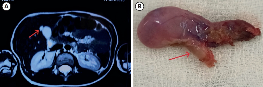
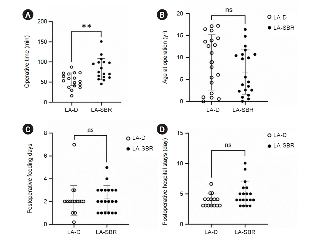
Purpose This study aimed to compare perioperative outcomes between laparoscopic-assisted diverticulectomy and laparoscopic-assisted small bowel resection for Meckel diverticulum.
Methods This single-center retrospective comparative cohort study included 39 patients who underwent laparoscopic-assisted surgery for Meckel diverticulum at Samsung Medical Center, Korea, between June 2010 and December 2023. Patients were classified into the laparoscopic-assisted diverticulectomy group or the laparoscopic-assisted small bowel resection group. Baseline characteristics, preoperative presentation, operative time, time to first oral feeding, postoperative hospital stay, complications, reoperation, and follow-up outcomes were compared between groups.
Results Of the 39 patients, 19 underwent laparoscopic-assisted diverticulectomy and 20 underwent laparoscopic-assisted small bowel resection. Baseline characteristics and preoperative presentation did not differ significantly between groups. Operative time was significantly shorter in the diverticulectomy group than in the small bowel resection group (median, 55.0 vs. 76.0 minutes; P=0.002). Time to first oral feeding did not differ significantly between groups (median, 2 days [2–2 days] vs. 2 days [1–3 days]; P=0.397). Postoperative hospital stays also did not differ significantly between groups (median, 4 days [3–4 days] vs. 4 days [4–5 days]; P=0.118), although hospitalization tended to be longer in the laparoscopic-assisted small bowel resection group. No statistically significant difference in complication rates was observed between groups; however, the number of events was low, limiting definitive comparison.
Conclusion Laparoscopic-assisted diverticulectomy was associated with shorter operative time and a tendency toward shorter postoperative hospital stay than small bowel resection, without an observed increase in early complications. It may be a reasonable option for carefully selected patients with uncomplicated Meckel diverticulum.
Benign cystic mesothelioma (BCM) is a rare intra-abdominal tumor and is particularly uncommon in pediatric patients. Its nonspecific clinical and radiological features often make preoperative diagnosis challenging. We report the case of a 4-year-old girl who presented with acute abdominal pain and vomiting. Computed tomography revealed a large, multiloculated cystic mass occupying the lower abdomen, which was initially suspected to be a lymphatic malformation. During laparoscopic exploration, a hemorrhagic, multiloculated cystic mass with hemoperitoneum was identified and completely resected with preservation of the right ovary. Histopathological examination confirmed BCM, showing predominant clusters of epithelioid cells interspersed with cyst-like spaces on hematoxylin and eosin staining. The patient recovered uneventfully and had no recurrence during 30 months of follow-up. This case describes an unusual presentation of BCM as an acute abdomen complicated by hemoperitoneum in a child and emphasizes the importance of surgical exploration and histopathological evaluation in establishing the diagnosis.
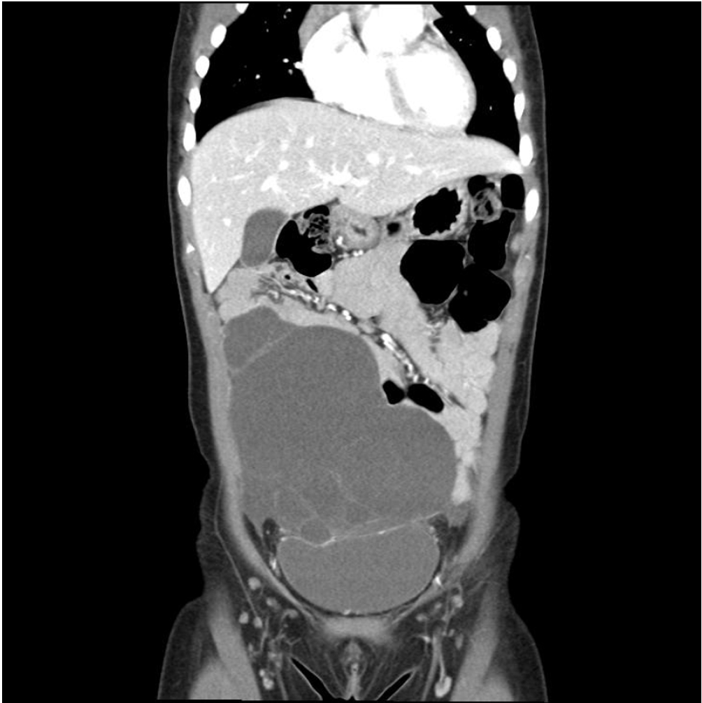
Purpose At our institution, congenital duodenal atresia is repaired laparoscopically using duodenoduodenostomy with a parallel anastomosis. During our use of this technique, we noted that after mobilization, the distal duodenal segment naturally rested in either a cranial or caudal orientation relative to the proximal segment and that this resting orientation appeared to vary with the anatomical level of atresia. This study primarily evaluated the association between atresia location and the natural orientation of the distal duodenal segment. It secondarily compared perioperative outcomes between the cranial and caudal orientation groups.
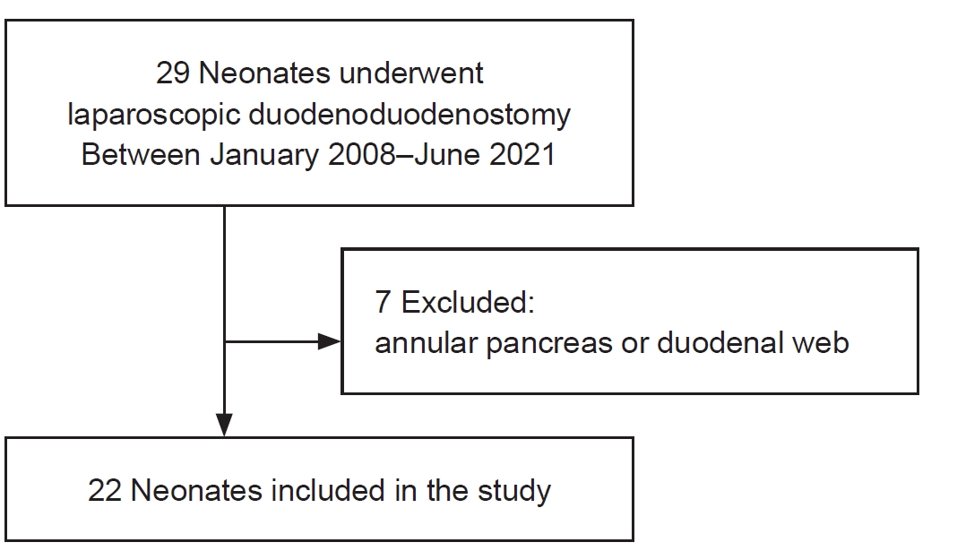
Methods This retrospective cohort study was conducted at Samsung Medical Center (Seoul, South Korea) and included neonates who underwent laparoscopic duodenoduodenostomy with parallel anastomosis for congenital duodenal atresia from January 2008 to June 2021. After patients with annular pancreas or duodenal web were excluded, 22 neonates were analyzed and categorized into the cranial (n=16) or caudal (n=6) orientation group according to intraoperative findings. Perioperative outcomes were compared, and the relationship between atresia location and distal segment orientation was analyzed.
Results Operative time, postoperative ventilator support, time to feeding initiation, time to full feeding, and length of hospitalization did not differ significantly between groups. No patient required conversion to open surgery, developed an anastomotic stricture, or died during hospitalization. One patient in the cranial group developed an anastomotic leak, which was treated by laparoscopic reanastomosis. First-portion duodenal atresia was significantly more frequent in the caudal group than in the cranial group (83.3% vs. 25.0%, P=0.023).
Conclusion Laparoscopic duodenoduodenostomy with parallel anastomosis was feasible in both cranial and caudal orientations, with no conversions to open surgery. The natural orientation of the distal duodenal segment was significantly associated with the anatomical location of atresia, supporting an anatomical basis for orientation-guided parallel anastomosis.


 First
First Prev
Prev

