1Department of Surgery, Ewha Womans University School of Medicine, Seoul, Korea.
2Department of Pathology, Ewha Womans University School of Medicine, Seoul, Korea.
Correspondence: Kum-ja Choi, M.D., Department of Surgery, Ewha Womans University School of Medicine, 911-1 Mok-dong, Yangcheon-gu, Seoul 158-710, Korea. Tel: 02)2650-5554, Fax: 02)2644-7984, kumchoi@ewha.ac.kr
• Received: September 26, 2012 • Accepted: November 15, 2012
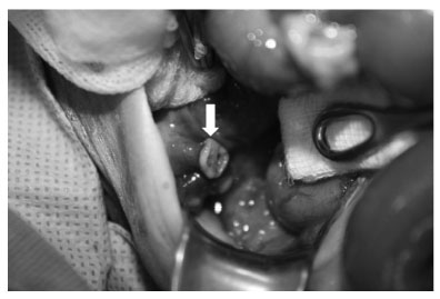
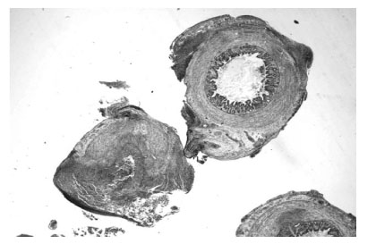
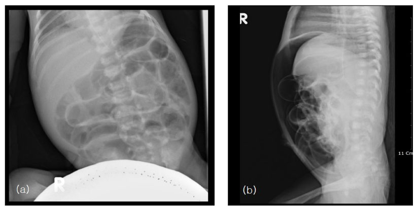
Acute appendicitis is very rare in premature neonates. Preoperative diagnosis of this condition is difficult, and then it leads to high morbidity and mortality. We report 9-day-old premature male with ruptured acute appendicitis presented with pneumoperitoneum on plain films of the abdomen. Awareness of this rare condition and possible differential diagnosis in this age group is also discussed.
12. Karaman A, Cavusoglu YH, Karaman I, Cakmak O. Seven cases of neonatal appendicitis with a review of the English language literature of the last century. Pediatr Surg Int. 2003. 19(11):707-709.
14. Khan TR, Rawat JD, Ahmed I, Rashid KA, Maletha M, Wakhlu A, Kureel SN. Neonatal pneumoperitoneum: a critical appraisal of its causes and subsequent management from a developing country. Pediatr Surg Int. 2009. 25:1093-1097.