ABSTRACT
Although Meckel's diverticulum is the most common vitellointestinal duct (VID) anomaly, patent vitellointestinal duct (PVID) is the most common symptomatic embryological defect. Patient may present with the anomaly itself or due to complications like intestinal obstruction secondary to volvulus, intussusception or adhesions. Prolapse occurs if the diverticulum is wide-mouthed enough to allow bowel to come out or due to increased intra-abdominal pressure like cry or cough. Bowel prolapse through PVID is rare and double prolapse of proximal as well as distal loop in a newborn is extremely rare. Omphalocele with prolapsing bowel through PVID as found in our index case is even rarer in literature. The pediatric surgeon should be familiar with these varied manifestations in the newborn because the prolapsed bowel can progress to gangrene and complications if not identified and operated upon early.
-
Keywords: Vitelline duct; Umbilical hernia; Ileal prolapse; Double prolapse
INTRODUCTION
Remnants of vitellointestinal duct (VID) account for a wide variety of umbilical abnormalities. These include fistula, sinus tract, umbilical adenoma, enterocystoma and congenital bands [
1]. Incidence of patent vitellointestinal duct (PVID) is reported as 0.0053% [
2]. Furthermore, bowel prolapse through PVID is rare and double prolapse of proximal as well as distal loop in a newborn is extremely rare. Omphalocele with prolapsing bowel through PVID as found in our index case is even rarer.
CASE REPORT
A full term male neonate, weighing 2,400 g, delivered by normal spontaneous vaginal delivery presented to our institute within 3 hours after birth; at first glance, he had a protruding mass through an umbilical defect covered with thin membrane and also had some prolapsed bowel loops. During the antenatal period, he was supervised with antenatal ultrasounds being normal. On examination, the cry, tone and activity of the baby were good. There was an omphalocele minor covered with a thin membrane and on the top of it were some prolapsed intestinal loops giving shape of an inverted Y (
Fig. 1). Meconium could be seen coming from the proximal limb of this Y. Patient was taken up for surgery after adequate hydration. Under general anesthesia, prolapsed loops were approached through circumferential incision and excision of omphalocele membrane. Intestine was delivered outside the abdomen and prolapsed bowel was reduced. A PVID opening was noted in the distal ileum at the site of prolapse (
Fig. 2). It was a wide mouthed PVID from where the ileal loops were intussuscepting and prolapsing outside. The intussuscepting bowel loop was gently reduced. The distal ileal segment with the PVID was resected and an end to end anastomosis of the bowel was done (
Fig. 3). The omphalocele had only distal part of ileum, entire cecum and ascending colon as its contents. These contents were replaced into the abdomen and umbilicoplasty was done with absorbable sutures (
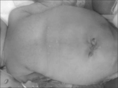
Fig. 4). Postoperative period was uneventful. Patient was started on small amount of feeds on 5th postoperative day, and discharged on full breast feeds on 13th postoperative day. At 3 months follow-up, the child is thriving well.
DISCUSSION
The VID connects the yolk sac to the gut in a developing embryo and provides nutrition until the placenta is established. Between the 5th and 7th week of gestation, the duct attenuates and separates from the intestine. Just before this involution, the epithelium of the yolk sac develops a lining similar to that of the stomach. Partial or complete failure of involution of the VID results in various residual structures. Meckel's diverticulum is the most common of these structures and is the most frequent congenital gastrointestinal anomaly occurring in 2% to 3% of all infants. Other VID remnants occur infrequently, including a persistently patent duct, a solid cord, or a cord with a central cyst or a diverticulum associated with a persistent cord between the diverticulum and the umbilicus [
34]. Bowel prolapse through PVID is rare and double prolapse of proximal as well as distal loop in a newborn is extremely rare [
56]. Furthermore, omphalocele with prolapsing bowel through PVID as seen in our index case is even rarer. After a thorough search of literature, we came across only five similar case reports [
7891011].
The condition, if not managed promptly by surgical intervention, may lead to subacute or acute intestinal obstruction, strangulation and gangrene of the prolapsed intestinal loops. Primary closure of the PVID following reduction of the prolapsed intestines may be possible if the patient arrives early without any gross edema over the intestinal loops. If the defect is large one can go for resection of the loop of intestine near the patent duct followed by primary anastomosis. If the patient arrives late with gross edema or the viability of the intestinal loops is in question then exteriorization of the suspected loop or loop ileostomy is advised [
12]. The good result with early primary resection and anastomosis in our case emphasizes the importance of prompt referral, early diagnosis and quick surgical intervention to prevent edema and gangrene which can be fatal. Even the morbidity associated with a stoma can be prevented if the pediatric surgeon is familiar with this unique presentation and decides to intervene early based on his clinical examination.
PVID with prolapsed ileal loop is a rare condition which needs prompt diagnosis, surgical reduction and repair of the defect. Since other malformations are not present, this entity may be regarded as a distinct entity, a form of a lower midline abdominal wall defect understood by a disturbance of development of vitelline duct and having excellent prognosis.
NOTES
-
No potential conflict of interest relevant to this article was reported.
REFERENCES
- 1. Cilley RE. Disorders of the umbilicus. In Grosfeld JL, O'Neill JA Jr, Coran AG, Fonkalsrud EW, editors. Pediatric surgery. 6th ed. St. Louis: Mosby/Elsevier; 2006, pp 1143-1156.
- 2. Chiang LS. Vitelline duct remnant appearing as a hemorrhagic umbilical mass. JAMA 1982;247:2812-2813.
- 3. Agrawal S, Memon A. Patent vitellointestinal duct. BMJ Case Rep 2010;doi: 10.1136/bcr.12.2009.2594
- 4. Ameh EA, Mshelbwala PM, Dauda MM, Sabiu L, Nmadu PT. Symptomatic vitelline duct anomalies in children. S Afr J Surg 2005;43:84-85.
- 5. Soutar SF, Douglas DM, Dennison WM. Patent vitello-intestinal duct; the risk of obstruction due to prolapse. Br J Surg 1958;45:617-620.
- 6. Borkar N. A prolapsing vitello-intestinal duct in newborn. APSP J Case Rep 2013;4:32.
- 7. Davis RM, Kehm RW. Omphalocele with patent vitellointestinal duct and ileal prolapse. Am J Surg 1967;113:571-573.
- 8. Rohatgi M, Gorthi SN. Omphalocele with patent omphalomesenteric duct and ileal prolapse. Indian J Pediatr 1984;51:119-123.
- 9. Storms P, Pexsters J, Vandekerkhof J. Small omphalocele with ileal prolapse through a patent omphalomesenteric duct. A case report and review of literature. Acta Chir Belg 1988;88:392-394.
- 10. Panait N, Michel F, D'Ercole C, Merrot T. Esophageal atresia, small omphalocele and ileal prolapse through a patent omphalomesenteric duct: a methamizole embryopathy? J Pediatr Surg 2013;48:E9-E11.
- 11. Blair SP, Beasley SW. Intussusception of vitello-intestinal tract through an exomphalos in trisomy 13. Ped Surg Int 1989;4:422-423.
- 12. Mohite PN, Bhatnagar AM, Hathila VP, Mistry JH. Patent vitellointestinal duct with prolapse of inverted loop of small intestine: a case report. J Med Case Rep 2007;1:49.
Fig. 1Umbilicus (asterisk), omphalocele (circle), site of prolapsed (rectangle) of patent vitellointestinal duct, proximal intussuscepted ileum (upwards arrow) from which some meconium was discharging at time of examination in emergency, distal intussuscepted ileum (downwards arrow).
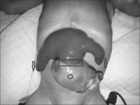
Fig. 2Intraoperative picture with arrow and forceps at site of wide mouthed patent vitellointestinal duct from where intussuscepting ileal loops were reduced.
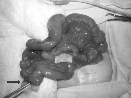
Fig. 3Intraoperative picture after resection and ileoileal end to end anastomosis at site of prolapsed patent vitellointestinal duct.
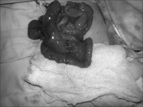
Fig. 4Postoperative picture shows well healed site of incision and umbilicoplasty.
Citations
Citations to this article as recorded by

- Omphalocele minor in a neonate with a patent vitellointestinal duct: a case report and insights on management from a systematic review of case reports
Rem Ehab Abdelkader, Lena Ehab Abdelkader, Joud Zghyer, Ehab Malek Abdelkader
Egyptian Pediatric Association Gazette.2026;[Epub] CrossRef - A rare case report of patent vitellointestinal duct with prolapsed orthograde ileal intussusception seen as bident horn
Jaya Ram Pandey, Prakash Kunwar, Suman Bikram Adhikari, Bal Mukund Basnet, Asmit Kumar Singh
Annals of Medicine & Surgery.2026; 88(6): 3667. CrossRef - Omphalocele with a patent vitelline duct: intraoperative identification of an uncommon presentation
Zoe Paige, Jay Kerecman, Sean Barnett
Maternal Health, Neonatology and Perinatology.2026;[Epub] CrossRef - Demographics, Clinical Presentation, and Surgical Procedures Performed for the Persistent Vitellointestinal Duct During Infancy: A Systematic Literature Review of the Past Fifty Years from 1971 to 2021
Rajendra K. Ghritlaharey
Medical Journal of Dr. D.Y. Patil Vidyapeeth.2024; 17(2): 262. CrossRef - Omphalocele with intestinal prolapse through a patent omphalomesenteric duct: A case report
SaraPettey Sandifer, Afif N. Kulaylat, Sara Mola, Aodhnait S Fahy
Journal of Pediatric Surgery Case Reports.2023; 98: 102722. CrossRef - Omphalocele with bladder prolapse through wide patent urachus
Tanvi Goel, Shilpa Sharma, Devasenathipathy Kandasamy, Minu Bajpai
Journal of Pediatric Surgery Case Reports.2023; 90: 102577. CrossRef - Bowel prolapse through umbilicus in newborn - an unforeseen emergency
Soumyodhriti Ghosh, Abhijit Kundu, Sunaram Majhi, Hari Ignatius Pandey, Abhishek Kumar
Pediatric Oncall.2023;[Epub] CrossRef - Exomphalos with intestinal fistulation: Case series and systematic review for clinical characterization, management and embryopathogenesis
Luke McNickle, Arjun Visa, Simon Clarke, Iain Yardley, Yew-Wei Tan
Journal of Pediatric Surgery.2022; 57(4): 661. CrossRef - Complete evagination of a patent vitellointestinal duct and adjacent ileal limbs from an omphalocele sac: an extreme presentation
Sarah Kher-ru Sim, Rambha Rai, Anette Sundfor Jacobsen
BMJ Case Reports.2019; 12(6): e229971. CrossRef - Occult Adenoma in Patent Vitellointestinal Duct Presenting as an Umbilical Fistula: Cause for Concern?
Himani Bhankar, Surbhi Goyal, Sufian Zaheer, Nidhi Sugandhi, Ashish Kumar Mandal
Fetal and Pediatric Pathology.2016; 35(4): 272. CrossRef


