1Department of Pediatric Surgery, Seoul National University Children's Hospital, Seoul, Korea.
2Department of Pediatric Surgery, Seoul National University College of Medicine, Seoul, Korea.
Correspondence: Hyun-Young Kim, Department of Pediatric Surgery, Seoul National University College of Medicine, 103 Daehak-ro, Jongno-gu, Seoul 03080, Korea. Tel: +82-2-2072-2478, Fax: +82-2-747-5130, spkhy02@snu.ac.kr
• Received: October 13, 2017 • Revised: October 25, 2017 • Accepted: October 25, 2017
This is an Open Access article distributed under the terms of the Creative Commons Attribution Non-Commercial License (https://creativecommons.org/licenses/by-nc/4.0) which permits unrestricted non-commercial use, distribution, and reproduction in any medium, provided the original work is properly cited.
Minimally invasive surgery (MIS) in abdomen and thorax has been widely accepted for pediatric diseases. Thoracoscopic surgery has the advantage of less pain, better cosmetic outcomes and less musculoskeletal sequelae in comparison to open surgery. We would like to share our initial experience with thoracoscopic surgery performed by one pediatric surgeon.
Methods
We performed a retrospective review of patients who underwent thoracoscopic surgery by one pediatric surgeon between April 2010 and August 2017 in Department of Pediatric Surgery, Seoul National University Children's Hospital.
Results
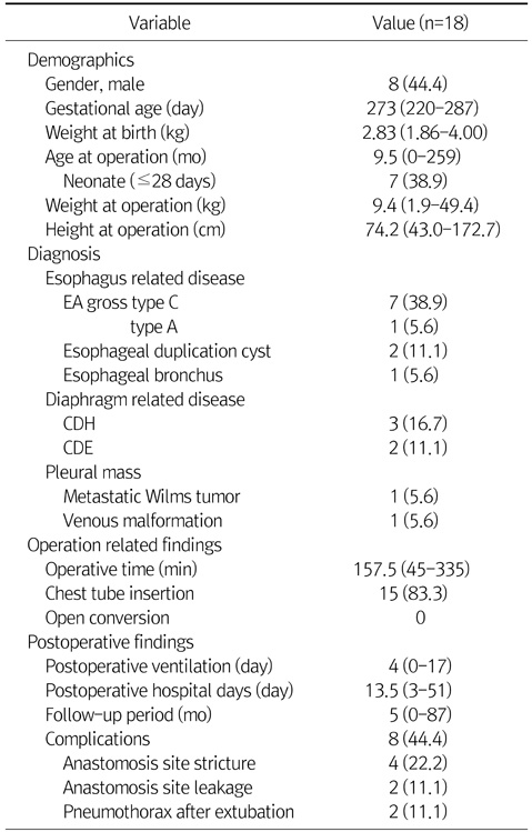
There were totally 18 cases; 8 cases for esophageal atresia, 3 cases for congenital diaphragm hernia, 2 cases for diaphragm eventration, 2 cases for esophageal duplication cyst, 2 cases for pleural mass and 1 case for esophageal bronchus. At the operation, median age was 9.5 months (range, 0-259 months) and median body weight was 9.4 kg (range, 1.9-49.4 kg). Median operative time was 157.5 minutes (range, 45-335 minutes). There was no case of open conversion and 2 cases of minor leakage at anastomosis site in case of esophageal atresia. Median follow-up month was 5 months (range, 0-87 months). During follow-up, 4 cases of esophageal atresia showed anastomosis site narrowing and average 2.5 times (range, 1-5 times) of esophageal balloon dilatation was done.
Conclusion
We performed thoracoscopic surgery in case of esophageal, diaphragm disease and pleural mass. Thoracoscopic surgery can be an effective and feasible option of treatment for well-selected pediatric patients of intra-thoracic disease including esophagus, diaphragm and mediastinum disease.
No potential conflict of interest relevant to this article was reported.
REFERENCES
1. Wall JK, Sinclair TJ, Kethman W, Williams C, Albanese C, Sylvester KG, et al. Advanced minimal access surgery in infants weighing less than 3kg: a single center experience. J Pediatr Surg 2017;05. 11. [Epub ahead of print].
2. Lieber J, Urla CI, Baden W, Schäfer J, Kirschner HJ, Fuchs J. Experiences and challenges of thorcoscopic lung surgery in the pediatric age group. Int J Surg 2015;23:169-175.
4. Yang YF, Dong R, Zheng C, Jin Z, Chen G, Huang YL, et al. Outcomes of thoracoscopy versus thoracotomy for esophageal atresia with tracheoesophageal fistula repair: a PRISMA-compliant systematic review and meta-analysis. Medicine (Baltimore) 2016;95:e4428.
5. Huang JS, Lau CT, Wong WY, Tao Q, Wong KK, Tam PK. Thoracoscopic repair of congenital diaphragmatic hernia: two centres' experience with 60 patients. Pediatr Surg Int 2015;31:191-195.
8. Zani A, Lamas-Pinheiro R, Paraboschi I, King SK, Wolinska J, Zani-Ruttenstock E, et al. Intraoperative acidosis and hypercapnia during thoracoscopic repair of congenital diaphragmatic hernia and esophageal atresia/tracheoesophageal fistula. Paediatr Anaesth 2017;27:841-848.
11. Holcomb GW 3rd, Rothenberg SS, Bax KM, Martinez-Ferro M, Albanese CT, Ostlie DJ, et al. Thoracoscopic repair of esophageal atresia and tracheoesophageal fistula: a multi-institutional analysis. Ann Surg 2005;242:422-428 discussion 428-30.
12. St Peter SD, Ostlie DJ. Laparoscopic gastric transposition with cervical esophagogastric anastomosis for long gap pure esophageal atresia. J Laparoendosc Adv Surg Tech A 2010;20:103-106.
14. Bratu I, Laberge JM, Flageole H, Bouchard S. Foregut duplications: is there an advantage to thoracoscopic resection? J Pediatr Surg 2005;40:138-141.
15. Fujishiro J, Ishimaru T, Sugiyama M, Arai M, Suzuki K, Kawashima H, et al. Minimally invasive surgery for diaphragmatic diseases in neonates and infants. Surg Today 2016;46:757-763.
16. Becmeur F, Reinberg O, Dimitriu C, Moog R, Philippe P. Thoracoscopic repair of congenital diaphragmatic hernia in children. Semin Pediatr Surg 2007;16:238-244.
17. Li B, Chen BW, Wang QS, Wang BY. Thoracoscopic repair of diaphragmatic hernia in infants: a new modification of the technique. Eur J Pediatr Surg 2016;26:519-523.
18. Clark RH, Hardin WD Jr, Hirschl RB, Jaksic T, Lally KP, Langham MR Jr, et al. Current surgical management of congenital diaphragmatic hernia: a report from the Congenital Diaphragmatic Hernia Study Group. J Pediatr Surg 1998;33:1004-1009.
19. Pierro A. Hypercapnia and acidosis during the thoracoscopic repair of oesophageal atresia and congenital diaphragmatic hernia. J Pediatr Surg 2015;50:247-249.
20. Yang EY, Allmendinger N, Johnson SM, Chen C, Wilson JM, Fishman SJ. Neonatal thoracoscopic repair of congenital diaphragmatic hernia: selection criteria for successful outcome. J Pediatr Surg 2005;40:1369-1375.
22. Kocaturk CI, Sezen CB, Aker C, Kalafat CE, Bilen S, Kutluk AC, et al. Surgical approach to posterior mediastinal lesions and long-term outcomes. Asian Cardiovasc Thorac Ann 2017;25:287-291.
23. Park T, Jung K, Kim HY, Jung SE, Park KW. Thoracoscopic management of a communicating bronchopulmonary foregut malformation in a 23-month-old child. J Pediatr Surg 2012;47:e21-e23.
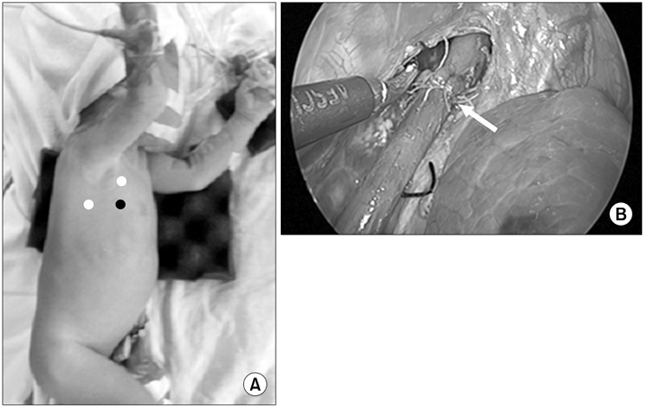
Esophageal atresia (EA) with tracheoesophageal fistula. (A) Position of patient for operation of EA (white dots, working port; black dot, camera port). (B) After ligating azygos vein, fistula was also ligated with simple suture, and end-to-end anastomosis of esophagus was done with interrupted suture. Arrow, end-to-end anastomosis site.
Fig. 2
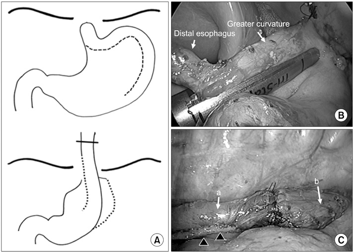
Esophageal atresia, Gross type A. (A) After 7 months, he underwent thoracoscopy and laparoscopy surgery simultaneously by gastric tube interposition. (B) Stomach was incised along great curvature with endo GIA, and gastric tube was made. (C) Gastric tube with distal esophagus was interpositioned to thorax, and esophagoesophagostomy was done by thoracoscopy. Arrows: a, gastric tube-distal part of esophagus; b, proximal part of esophagus. Arrowheads, resected with Endo GIA (Covidien, USA).
Fig. 3
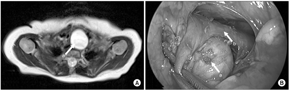
Esophageal duplication cyst located at posterior mediastinum. (A) The mass was abutting with anterior wall of esophagus on chest CT (2.5×1.9×2.5-cm-sized mass). (B) Mass was connected to muscle layer of esophagus. Arrows: a, mass; b, esophagus.
Fig. 4
Esophageal duplication cyst of superior mediastinum. (A) There was a mass situated at just front of upper esophagus on neck MRI (2.5×2.2×4.5-cm-sized mass). (B) There was a mass at the front and right of the esophagus. Arrows: a, esophageal duplication cyst; b, true esophagus.
Fig. 5
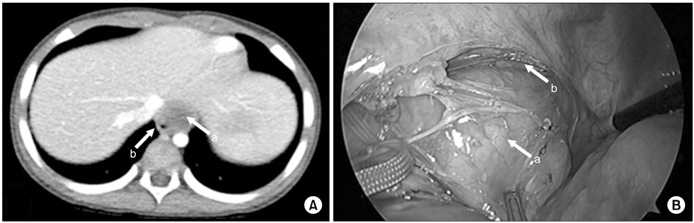
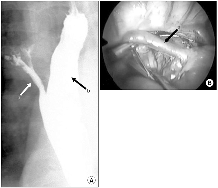
Esophageal bronchus. (A) Upper gastrointestinal series showed a community between esophagus and trachea. The contrast also showed that bronchus and bronchioles are risen from esophagus. (B) Intraoperative photo. There was fistula tract between bronchus and esophagus, so it was ligated with simple suture. Patient underwent thoracoscopic right upper lobectomy and fistula division. Arrows: a, fistula tract; b, esophagus.
Fig. 6
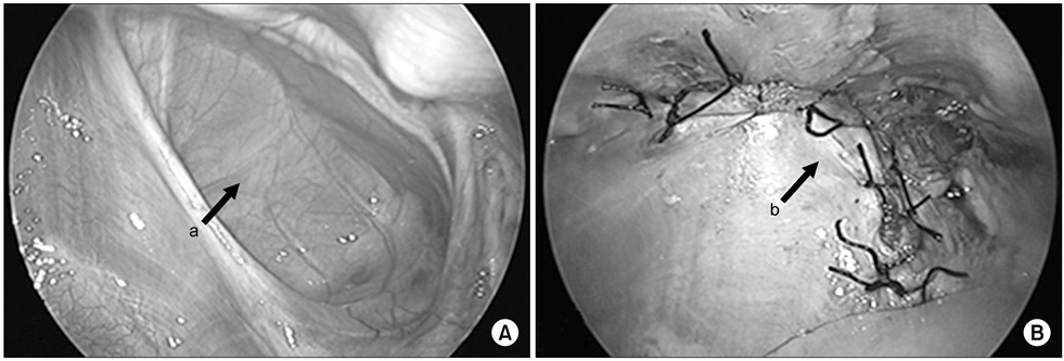
Congenital diaphragm hernia. (A) There was hernia sac on right diaphragmatic defect. (B) Hernia sac was excised and primary repair was performed. Arrows: a, hernia sac; b, primary closure.
Fig. 7
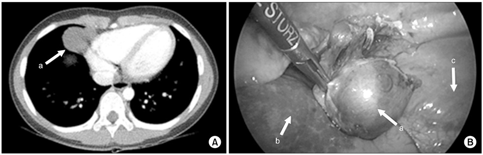
Venous malformation. (A) There was mass at cardoiphrenic angle and it was suspected an inflammatory pseudo-tumor or congenital lung lesion like cystic lymphangioma on chest CT (3.0×3.6×2.3-cm-sized mass). (B) It was being originated from right pleura and was resected clearly. Arrows: a, mass; b, lung; c, diaphragm.
Early Experience of Pediatric Thoracoscopic Surgery Performed by a Pediatric Surgeon
Fig. 1
Esophageal atresia (EA) with tracheoesophageal fistula. (A) Position of patient for operation of EA (white dots, working port; black dot, camera port). (B) After ligating azygos vein, fistula was also ligated with simple suture, and end-to-end anastomosis of esophagus was done with interrupted suture. Arrow, end-to-end anastomosis site.
Fig. 2
Esophageal atresia, Gross type A. (A) After 7 months, he underwent thoracoscopy and laparoscopy surgery simultaneously by gastric tube interposition. (B) Stomach was incised along great curvature with endo GIA, and gastric tube was made. (C) Gastric tube with distal esophagus was interpositioned to thorax, and esophagoesophagostomy was done by thoracoscopy. Arrows: a, gastric tube-distal part of esophagus; b, proximal part of esophagus. Arrowheads, resected with Endo GIA (Covidien, USA).
Fig. 3
Esophageal duplication cyst located at posterior mediastinum. (A) The mass was abutting with anterior wall of esophagus on chest CT (2.5×1.9×2.5-cm-sized mass). (B) Mass was connected to muscle layer of esophagus. Arrows: a, mass; b, esophagus.
Fig. 4
Esophageal duplication cyst of superior mediastinum. (A) There was a mass situated at just front of upper esophagus on neck MRI (2.5×2.2×4.5-cm-sized mass). (B) There was a mass at the front and right of the esophagus. Arrows: a, esophageal duplication cyst; b, true esophagus.
Fig. 5
Esophageal bronchus. (A) Upper gastrointestinal series showed a community between esophagus and trachea. The contrast also showed that bronchus and bronchioles are risen from esophagus. (B) Intraoperative photo. There was fistula tract between bronchus and esophagus, so it was ligated with simple suture. Patient underwent thoracoscopic right upper lobectomy and fistula division. Arrows: a, fistula tract; b, esophagus.
Fig. 6
Congenital diaphragm hernia. (A) There was hernia sac on right diaphragmatic defect. (B) Hernia sac was excised and primary repair was performed. Arrows: a, hernia sac; b, primary closure.
Fig. 7
Venous malformation. (A) There was mass at cardoiphrenic angle and it was suspected an inflammatory pseudo-tumor or congenital lung lesion like cystic lymphangioma on chest CT (3.0×3.6×2.3-cm-sized mass). (B) It was being originated from right pleura and was resected clearly. Arrows: a, mass; b, lung; c, diaphragm.
Fig. 1
Fig. 2
Fig. 3
Fig. 4
Fig. 5
Fig. 6
Fig. 7
Early Experience of Pediatric Thoracoscopic Surgery Performed by a Pediatric Surgeon