Department of Surgery, Yeouido St. Mary's Hospital, School of Medicine, The Catholic University of Korea, Seoul, Korea.
Copyright © 2016 by the Korean Association of Pediatric Surgeons
This is an Open Access article distributed under the terms of the Creative Commons Attribution Non-Commercial License (https://creativecommons.org/licenses/by-nc/4.0) which permits unrestricted non-commercial use, distribution, and reproduction in any medium, provided the original work is properly cited.
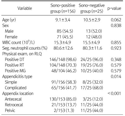
Values are presented as mean±SD or n (%).
Seg., segmented; exam., examination; RLQ, right lower quadrant; DT, direct tenderness; RT, rebound tenderness; MG, muscle guarding.
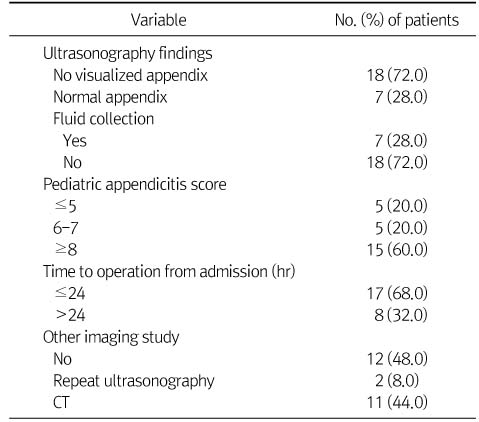
Values are presented as mean±SD or n (%).
Seg., segmented; exam., examination; RLQ, right lower quadrant; DT, direct tenderness; RT, rebound tenderness; MG, muscle guarding.