1Department of Pediatric Surgery, Seoul National University Children's Hospital, Seoul, Korea.
2Department of Pediatric Surgery, Seoul National University College of Medicine, Seoul, Korea.
Copyright © 2018 Korean Association of Pediatric Surgeons
This is an Open Access article distributed under the terms of the Creative Commons Attribution Non-Commercial License (https://creativecommons.org/licenses/by-nc/4.0) which permits unrestricted non-commercial use, distribution, and reproduction in any medium, provided the original work is properly cited.
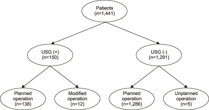


Values are presented as mean±SD or n (%).
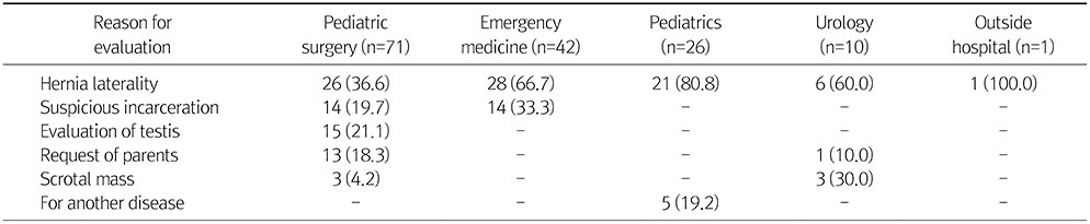
Values are presented as n (%).
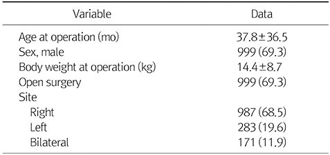
Values are presented as mean±SD or n (%).
Values are presented as n (%).