

Purpose
Abdominal computed tomography (ACT) is widely used to diagnose appendicitis in children. Despite its high sensitivity and specificity, “negative appendectomies” still occur when the patient undergoes surgery but the final pathologic diagnosis does not support appendicitis. The aim of this study is to determine which findings support true appendicitis in patients with unclear findings on preoperative ACT.
Methods
We performed a retrospective review of the records of 620 pediatric patients who underwent surgery for acute appendicitis between January 1, 2007 and December 31, 2020.
We re-reviewed the scans in 101 patients who were deemed to have unclear preoperative findings on ACT, looking for the following features: periappendiceal fat infiltration, periappendiceal fluid collection, appendiceal wall thickening, appendiceal gas, and right lower quadrant lymphadenopathy. We then compared the presence of these features between patients with true appendicitis and those who underwent negative appendectomy.
Results
The presence of an enlarged appendix, with a maximum diameter of more than 8 mm, and the presence of periappendiceal fat infiltration were associated with true appendicitis.
Conclusion
If ACT findings are unclear in a patient with suspected acute appendicitis, the presence of an enlarged appendix and periappendiceal fat infiltration should be assessed to differentiate those with true appendicitis.
Abdominal computed tomography (ACT) is widely used to diagnose appendicitis in children. Despite its high sensitivity and specificity, “negative appendectomies” still occur when the patient undergoes surgery but the final pathologic diagnosis does not support appendicitis. The aim of this study is to determine which findings support true appendicitis in patients with unclear findings on preoperative ACT.
We performed a retrospective review of the records of 620 pediatric patients who underwent surgery for acute appendicitis between January 1, 2007 and December 31, 2020. We re-reviewed the scans in 101 patients who were deemed to have unclear preoperative findings on ACT, looking for the following features: periappendiceal fat infiltration, periappendiceal fluid collection, appendiceal wall thickening, appendiceal gas, and right lower quadrant lymphadenopathy. We then compared the presence of these features between patients with true appendicitis and those who underwent negative appendectomy.
The presence of an enlarged appendix, with a maximum diameter of more than 8 mm, and the presence of periappendiceal fat infiltration were associated with true appendicitis.
If ACT findings are unclear in a patient with suspected acute appendicitis, the presence of an enlarged appendix and periappendiceal fat infiltration should be assessed to differentiate those with true appendicitis.
Acute appendicitis is the most common abdominal surgical emergency in pediatric patients. Despite its high incidence, diagnosing acute appendicitis is often a challenge [1]. The classic presenting symptoms—such as periumbilical pain, nausea, vomiting, and migration of pain to the right lower quadrant—are not always reported and are less reliable in children, particularly those younger than 5 years of age [2, 3, 4]. Imaging plays a key role in the accurate and prompt diagnosis of suspected appendicitis when the clinical presentation is equivocal [5, 6]. As an initial imaging modality for suspected acute appendicitis, ultrasonography (USG) has been shown to have high diagnostic accuracy, reducing or obviating the need for further imaging studies. While USG is highly accurate, it is operator-dependent and affected by patient-specific factors, including obesity [7, 8, 9]. Because of its speed and accuracy, abdominal computed tomography (ACT) is currently in widespread use in pediatric patients who have an equivocal clinical presentation [10, 11, 12]. Although ACT has a higher sensitivity and specificity than USG, somewhere between 3.6% and 6.7% of patients still end up undergoing “negative appendectomy”, with surgical pathology revealing a benign appendix [13].
Radiologists interpreting the results of ACT divide the scans into 5 categories, using Stengel's classification system: grade 1, definitely not appendicitis; grade 2, non-visualization of the appendix with no secondary signs of inflammation; grade 3, equivocal; grade 4, probable appendicitis; and grade 5, high probability of appendicitis. Using this classification system, only a grade of 1 or 5 actually helps surgeons to make a clear decision about surgery [14].
The purpose of this study is to elucidate the findings on ACT that support true appendicitis in patients with preoperative ACT grades 2, 3, and 4 (the unclear grades) by comparing the ACT findings between patients with pathologically confirmed appendicitis and those with benign pathology. We hope that our findings can help patients and surgeons avoid unnecessary surgery.
The first-line imaging modality for appendicitis was ACT, and no further work-up was routinely considered for suspected cases. We performed a retrospective review of the medical records of 620 pediatric patients who underwent surgery for acute appendicitis between January 1, 2007 and December 31, 2020. We then excluded patients who underwent only USG or who had no imaging performed before surgery. We also excluded patients whose preoperative ACT results were classified as grade 1 or grade 5 according to Stengel's classification. We took the remaining patients—those who underwent appropriate preoperative imaging and had a Stengel's classification of 2, 3, or 4—and divided them into 2 groups according to their pathology report; this gave us our “true appendicitis” group and our “negative appendicitis” group.
The ACT images were retrospectively assessed for the following features: appendiceal enlargement, presence of an appendicolith or hyperdense material in the appendix, periappendiceal fat inflammation, periappendiceal fluid collection, appendiceal wall thickening, appendiceal gas, and right lower quadrant lymphadenopathy. As the Stengel's original classification was based the observations on adult cases, we had modified the reference values according to the observations on pediatric cases and as follow. The diameter of the appendix was measured from outer wall to outer wall; the appendix was considered enlarged if the diameter was greater than 8 mm [15]. An appendicolith was defined as an intraluminal lesion of high density (similar to that of the adjacent bone) [16]. Hyperdense material in the appendix was defined as an intra-appendiceal lesion that appeared denser than the adjacent bowel wall [17]. Appendiceal wall thickness was measured from the luminal surface to the serosal surface and was considered to be thickened if it measured more than 2.2 mm [18]. Lymphadenopathy was defined as a lymph node measuring greater than 8 mm at its smallest diameter [19].
All measured features were analyzed as dichotomous variables. Statistical analyses were performed using SPSS software, version 27.0 (SPSS Inc., Chicago, IL, USA). Dichotomous variables were compared using the χ2 test between the true appendicitis and negative appendicitis groups. Statistical significance was defined as a p-value less than 0.05.
This study was approved by the Kangwon National University Hospital Institutional Review Board (KNUH-2015-08-003).
Of the 620 pediatric patients who underwent appendectomy during the study period, we excluded those who underwent only USG (n=108) or who had no imaging performed (n=30) prior to surgery. We also excluded patients whose preoperative ACT results were classified as grade 1 (n=15) or grade 5 (n=366) according to Stengel's classification. A total of 101 patients underwent appropriate preoperative imaging and had a Stengel's classification of 2, 3, or 4. We divided these patients into 2 groups according to their pathology results: the true appendicitis group (n=88) and the negative appendicitis group (n=13).
Of the original 620 appendectomies performed, 53 were considered “negative appendectomies”. The overall negative appendectomy rate at our institution was 8.5%. In the study group (n=101), 13 patients had a negative appendectomy, giving a negative appendectomy rate of 12.9%. There was no statistical difference between the negative and true appendicitis groups regarding patient age or sex. One patient in the negative appendicitis group underwent ACT after an unclear USG. Most patients in the true appendicitis group had simple appendicitis, but the negative appendicitis group had a variety of conditions noted, including serosal congestion, lymphoid hyperplasia, and submucosal lipomatosis. The pathological results are summarized in Table 1.
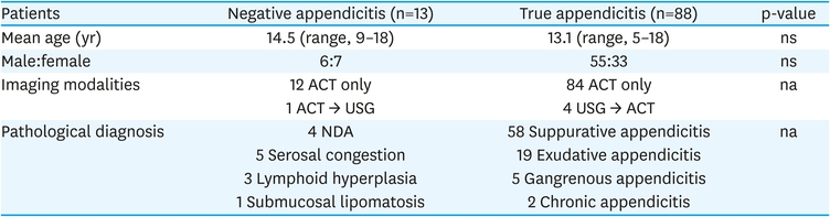
Table 1
Patient demographics
The mean maximal diameter of the appendix was 9.56 mm in the true appendicitis group and 7.28 mm in the negative appendicitis group. According to the definition of appendiceal enlargement (>8 mm in maximal diameter), 3 patients (23.1%) in the negative appendicitis group had an enlarged appendix, while 60 patients (68.2%) in the true appendicitis group had appendiceal enlargement (Fig. 1). This difference was statistically significant (p=0.002). Periappendiceal fat inflammation, or fat stranding, was noted in 2 patients (15.4%) in the negative appendicitis group and 43 patients (48.9%) in the true appendicitis group (p=0.023) (Fig. 2). For the other measured findings, there were no significant differences between the groups. The ACT findings are summarized in Table 2, along with their odds ratios and 95% confidence intervals (CIs).
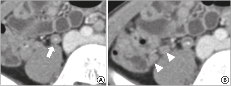
Fig. 1
Preoperative abdominal computed tomography findings for a 9-year-old male patient, who was demonstrated to have serositis: (A) appendicolith with focal wall thickening (arrow) and (B) adjacent mesenteric lymphadenopathy (arrowheads) were observed, but there was no associated periappendiceal fat infiltration or appendiceal enlargement.
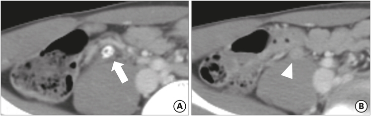
Fig. 2
Preoperative ACT findings for a 13-year-old male patient, who was demonstrated to have lymphoid hyperplasia: (A) appendicolith (arrow) and (B) luminal distension (arrowheads) were observed, but there were no other diagnostic abnormalities in ACT.
ACT, abdominal computed tomography.
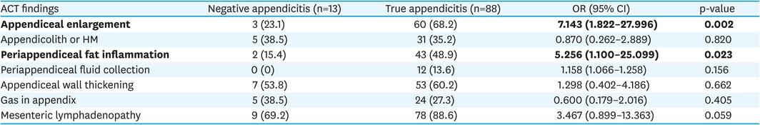
Table 2
Comparison of ACT findings between negative and true appendicitis groups
It is challenging to accurately diagnose acute appendicitis in pediatric patients who present with abdominal pain. In the past, appendicitis was diagnosed based on the clinical presentation and laboratory results, and the negative appendectomy rate approached 20% [20]. With the incorporation of imaging into diagnosis, the negative appendectomy rate decreased to around 8.5% [21]. It is important to note that USG is operator-dependent, and there are limitations to its use, including noncooperation in young patients, patient characteristics such as obesity, and anatomical diversity in the position of the appendix. Although the use of ACT has issues, including radiation exposure and the potential for contrast allergy, it is frequently performed due to its ability to provide rapid, accurate results [22].
During the study period, the rate of negative appendectomy at our institution was 8.5%, a rate similar to that noted at other institutions. However, the rate of negative appendectomy increased to 12.9% in patients with unclear ACT results—the patients in our study group. This means that a more refined approach to interpretation of ACT results is needed in patients with unclear results (grades 2, 3, and 4).
The ACT diagnosis of acute appendicitis is typically based on visualization of an appendix greater than 6 mm in maximal diameter, with contrast enhancement in the thickened appendiceal wall or pericecal inflammatory changes. The diagnosis can also be made when a phlegmon or abscess is visualized, with or without an appendicolith [18]. Bursali et al. [23] report that the outer-wall diameter of a normal appendix does not exceed 6 mm, and Ives et al. [24] report that the maximal outer-wall diameter of a normal appendix averages 6.6 mm (±1.0 mm). However, Taylor et al. [11] state that an appendix enlarged to greater than 6 mm in diameter is not sufficient for the diagnosis of acute appendicitis. In our study, we used a cutoff of 8 mm to define an enlarged appendix in patients with unclear ACT results. In such patients, it is not necessary to determine whether the appendix is normal or not, but rather to distinguish acute appendicitis from a benign process. Setting the definition of enlargement at 8 mm improves the accuracy of diagnosis. In our negative appendicitis group, the average appendiceal diameter was 7.28 mm; in the true appendicitis group, the average appendiceal diameter was 9.53 mm. Periappendiceal infiltration (fat stranding) is usually of value as a secondary sign of acute appendicitis, according to Rao et al. [20]. We noted that fat infiltration was significantly different between our true appendicitis and negative appendicitis groups.
Talyor et al. [11] conclude that an isolated appendicolith noted on ACT without signs of inflammation or with minimal fat stranding is not sufficient to diagnose acute appendicitis. We noted that 38.5% of our negative appendicitis group had an appendicolith or hyperdense material within the appendix, and none of the patients in the negative appendicitis group had a fluid collection visible on ACT. Therefore, in patients with an appendicolith but without any secondary signs of appendicitis, clinicians need to think of other diagnoses.
To reduce the radiation exposure in younger patients, the American College of Radiology recommends a staged protocol, using first USG and then ACT (follow-up ACT is recommended when the USG findings are equivocal). Krishnamoorthi et al. [25] found that using a staged USG-ACT protocol is as good as ACT alone in terms of sensitivity and specificity for acute appendicitis. Although the use of this staged protocol has reduced the use of ACT, the latter is still being performed in a large number of patients. Our findings could help to make optimal use of this imaging modality, leading to a more accurate diagnostic capability.
Magnetic resonance imaging (MRI) has a sensitivity and specificity similar to that of ACT. Therefore, MRI is an alternative modality that can reduce radiation exposure and prevent contrast-induced allergic reactions and nephrotoxicity; this is particularly useful in pediatric and pregnant patients [26]. Although we were not able to make use of MRI for the diagnosis of appendicitis due to the limitations of our national payment system, further studies are needed to clarify the role of MRI when there is diagnostic uncertainty.
Benabbas et al. [27] conclude that the factors with the strongest association with acute appendicitis in patients with undifferentiated abdominal pain are a history of pain migration to the right lower quadrant (positive likelihood ratio [LR+], 4.81; 95% CI, 3.59–6.44) and the presence of “cough/hop pain” on physical examination (LR+, 7.64; 95% CI, 5.94–9.83). Kwan et al. [28] found that the combination of a leukocyte count of 12,000/mm3 or greater and a C-reactive protein level greater than 3 mg/dL have the highest LR+ (4.36; 95% CI, 2.26–8.42) in patients with undifferentiated acute abdominal pain; however, this combination of laboratory results is only described in a single study. Minneci et al. [29] show that nonoperative management with antibiotics has a success rate of 67.1% in patients with uncomplicated acute appendicitis. In patients with suspected uncomplicated appendicitis based on physical findings, laboratory results, and unclear ACT results, a management strategy that includes the use of antibiotics with close follow-up is another good tool to reduce the rate of negative appendectomy.
In conclusion, certain findings on ACT are suggestive of true vs negative appendicitis in pediatric patients with unclear ACT results. The presence of an enlarged appendix (>8 mm in diameter) and the presence of periappendiceal fat inflammation are strongly associated with true appendicitis. These parameters can be useful for children with unclear ACT findings in whom appendicitis is suspected, helping to decrease the rate of negative appendectomy.
Conflict of Interest:No potential conflict of interest relevant to this article was reported.
Author Contributions:
Conceptualization: M.S.B.
Data curation: S.B., M.S.B., C.S.J., C.G., P.S.B., H.S.K., K.Y.H., K.H.
Formal analysis: S.B., M.S.B.
Investigation: S.B., M.S.B.
Methodology: S.B., M.S.B.
Supervision: M.S.B.
Validation: M.S.B.
Writing - original draft: S.B.
Writing - review & editing: M.S.B.
