

Sigmoid volvulus (SV) occurs due to the twist of a dilated sigmoid colon on its mesenteric axis, which can compromise the blood supply to the colon, leading to necrosis or perforation of the sigmoid colon. Potentially life threatening, SV is common in the elderly and rare in youth. We present the case of a 16-year-old boy who had experienced 3 episodes of SV over the course of a year and was successfully treated with laparoscopic-assisted sigmoid colectomy. SV should be considered when a young patient has a history of recurrent abdominal pain, constipation, and abdominal distension.
Sigmoid volvulus (SV) occurs due to the twist of a dilated sigmoid colon on its mesenteric axis, which can compromise the blood supply to the colon, leading to necrosis or perforation of the sigmoid colon. Potentially life threatening, SV is common in the elderly and rare in youth. We present the case of a 16-year-old boy who had experienced 3 episodes of SV over the course of a year and was successfully treated with laparoscopic-assisted sigmoid colectomy. SV should be considered when a young patient has a history of recurrent abdominal pain, constipation, and abdominal distension.
Sigmoid volvulus (SV) occurs due to the twist of a dilated sigmoid colon on its mesenteric axis, causing an obstruction and compromising the blood supply to the colon. Impaired blood supply can cause necrosis or perforation of the sigmoid colon [1, 2]. If left untreated, SV can result in a potentially life-threatening perforation [2, 3]. As a person ages, the S-shaped part of the colon or the part attached to the abdominal wall can stretch out, allowing it to twist around itself, resulting in a volvulus [4]. Thus, SV is common in the elderly and rare in children and youth. SV is considered a medical emergency and physicians should first attempt to reduce the SV. Subsequently, definitive surgery should be performed due to high rates of recurrence (55%–90%) [5]. Herein, we present the case of a 16-year-old boy who had 3 episodes of SV and was successfully treated with laparoscopic-assisted sigmoid colectomy.
A previously healthy 16-year-old boy with stable vital signs presented with vomiting, diarrhea, distension, and abdominal pain for 4 days. On physical examination, he had abdominal distension with mild abdominal tenderness but without rebound tenderness, and a subtle metallic bowel sound. Laboratory test results were normal. An abdominal radiography revealed a dilated sigmoid colon on the left quadrant (Fig. 1). An abdominal computed tomography (CT) revealed a marked distension of the gas-filled sigmoid colon with twisting of the mesenteric vessels, confirming SV (Fig. 2). A rectal tube was inserted to decompress the volvulus (Fig. 3). He remained asymptomatic thereafter, and discharge was requested. At 2 and 7 months after his initial attack, he was readmitted to the emergency department with the same complaints. Subsequent examinations confirmed the recurrence of SV. The SV was successfully decompressed by means of a rectal tube. His parents again refused surgical treatment; however, 9 months after the first attack, he underwent laparoscopic-assisted sigmoid colectomy. We use a 12 mm port above the umbilicus, a 5 mm right upper quadrant port, a 5 mm left sided port, and a 12 mm right lower quadrant port & left lower quadrant, typically placed 2 fingerbreadths medial and 2 fingerbreadths cephalad to the anterior superior iliac spine. After mobilization of the sigmoid colon, the colon was transected at the rectosigmoid junction with an endolinear stapler (SigniaTM Stapling System; Medtronic, Minneapolis, MN, USA). The umbilical incision was extended. The divided sigmoid colon was exteriorized, and the redundant part was resected. A sigmoid colon section of 34 cm was resected; end-to-end colorectal anastomosis was performed intracorporeally using a circular stapler (ECHELON CIRCULARTM 25 mm Powered Stapler; Ethicon, Bridgewater, NJ, USA). A seromuscular biopsy specimen from the distal colon submitted for histopathologic examination showed normal ganglion cells. On postoperative day 3, he was started on water intake; subsequently, eating progressed successfully. He was discharged without complications on postoperative day 10.
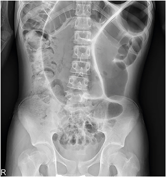
Fig. 1
Abdominal X-ray showing a dilated sigmoid colon on the left quadrant with the characteristic coffee bean sign.
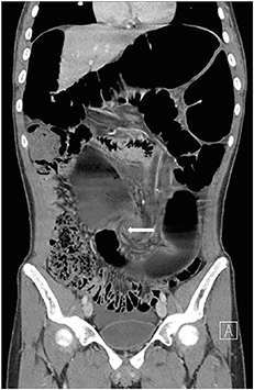
Fig. 2
Coronal abdominal computed tomography showing a dilated sigmoid colon with whirl pattern (arrow) of the mesentery.
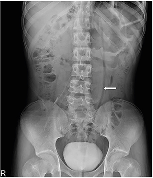
Fig. 3
Abdominal X-ray showing decompression of the volvulus by means of a rectal tube (arrow).
The colon is a part of the alimentary tract commonly affected by volvulus with the most common site of volvulus being the sigmoid colon [6]. The etiology of SV is multifactorial, including important predisposing factors, such as chronic constipation, high fiber diet, male sex, bowel habits, high altitude, ginger enema, as well as sigmoid colonic length [7, 8]. The length and width of the sigmoid colon, as well as those of the mesocolon, changes as patients' age, and the largest dimension change occurs in the elderly population [9]. Elongation and dilatation of the sigmoid colon may develop following constipation and fecal impaction or frequent distension of the colon by bulky stools in patients eating a high-fiber diet, thereby explaining that SV is relatively more frequent in the elderly [7]. In contrast to adults, the etiology of SV in children is believed to involve a pathologically long and redundant colon and a narrow-based mesentery or lack of fixation of a normally fixed part of the colon [10]. Predisposing factors in children include a history of imperforate anus, prune belly syndrome, intestinal malrotation, Hirschsprung's disease, and mental retardation [5].
Two different clinical presentations have been reported in children, depending on the age of the patient [5]. Children under 10 years of age usually experience an acute episode of abdominal pain for several hours without previous complaints. By the second decade of life, however, the picture of SV is that of a chronic disease with recurrent episodes of abdominal pain, constipation for several days, nausea, abdominal distension, and occasional vomiting; i.e., usually self-limited symptoms that subside following the passage of flatus and feces [5, 11, 12].
Due to spontaneous reduction of the volvulus in the second decade of life, a diagnosis may be missed or delayed [5, 11]. Many of these patients are diagnosed with irritable bowel syndrome, chronic constipation, or chronic abdominal pain [12]. Delayed diagnosis of SV in children is associated with high mortality (21%–29%) [13]. The coffee bean sign is a classic conventional radiographic finding of volvulus. However, the sensitivity of the coffee bean sign for the SV in children is reported to be only 16%–29% when reviewing pediatric cases in the literature [5, 14]. Dilated large bowel proximal to the sigmoid and air-fluid levels in the small bowel loops are often present in patients with SV, but can also be seen in patients with other causes of distal colonic obstruction, acute colonic pseudo-obstruction, constipation, or toxic megacolon [5]. The use of contrast enema with a water-soluble contrast can help diagnose a SV patient without evidence of peritonitis on physical examination, when abdominal radiographs are not definitive [5]. In cases where the diagnosis is doubtful, an abdominal CT scan may be a useful adjunctive modality. CT has proven useful for identifying the cause and site of obstruction resulting from other pathologies and for demonstrating ischemia caused by strangulation [15].
The goal of SV treatment is to decompress the bowel and prevent recurrent attacks. If there is no colonic necrosis or perforation, bowel decompression can initially be performed conservatively using a rectal tube, barium enema, or endoscopy [14]. The recurrence rate for SV with only non-operatively treatment is high [16, 17, 18]. In our case, the patient had experienced 3 recurrent attacks of SV. In the first episode, he visited the hospital after experiencing symptoms associated with SV for 4 days. In the subsequent 2 relapses, however, he visited the hospital on the day of symptom onset and was treated with rectal tube insertion immediately after diagnosis. Although definite treatment had not been performed on several attacks of SV, it is thought that prompt reduction treatment prevented the progression of intestinal necrosis. Johansson et al have reported that the recurrence rate following successful non-operative decompression at a second episode of SV was increased to 87.9% in contrast to the recurrence rate of 78.4% after the first episode [18]. Therefore, even in a patient suffering from the first attack of SV, surgery should be considered.
SV is rare in children; however, the true incidence is probably higher, due to spontaneous resolution. SV should be considered when a young patient has a history of recurrent abdominal pain, constipation, and abdominal distension that resolves by the passing of gas and stool. The importance of early diagnosis and surgical resection is crucial in preventing higher morbidity and mortality associated with recurrence.
Conflict of Interest:No potential conflict of interest relevant to this article was reported.
Author Contributions:
Conceptualization: K.K.H.
Data curation: C.S.Y.
Supervision: K.K.H.
Writing - original draft: C.S.Y.
Writing - review & editing: K.K.H.
