1Department of Pediatric Surgery, Donsgan Medical Center, Keimyung University School of Medicine, Daegu, Korea.
2Department of Pathology, Donsgan Medical Center, Keimyung University School of Medicine, Daegu, Korea.
Copyright © 2016 by the Korean Association of Pediatric Surgeons
This is an Open Access article distributed under the terms of the Creative Commons Attribution Non-Commercial License (https://creativecommons.org/licenses/by-nc/4.0) which permits unrestricted non-commercial use, distribution, and reproduction in any medium, provided the original work is properly cited.
The content of this article was presented at the 30th Annual Meeting of the Korean Association of Pediatric Surgeons, Busan, June 2014.
No potential conflict of interest relevant to this article was reported.
Sensitivity 100%, specificity 100%, positive predictive value 100%, negative predictive value 100%, accuracy 100%, p<0.001.
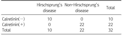


Sensitivity 100%, specificity 100%, positive predictive value 100%, negative predictive value 100%, accuracy 100%, p<0.001.
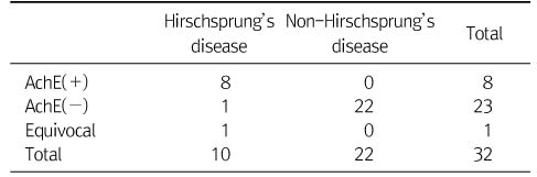
Sensitivity 80%, specificity 100%, p<0.001.
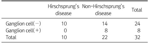
p=0.035.
Sensitivity 100%, specificity 100%, positive predictive value 100%, negative predictive value 100%, accuracy 100%, p<0.001.
Sensitivity 80%, specificity 100%, p<0.001.
p=0.035.