

Purpose
Currarino syndrome is a rare disease, and understanding its clinical characteristics is important because it involves complex anomalies and it requires a multidisciplinary approach for treatment. However, the accumulation of treatment experiences is challenging, and studies on this disease remain insufficient. Our study aimed to review the clinical characteristics and treatment of Currarino syndrome at our institution.
Methods
The medical records of patients diagnosed as Currarino triad or Currarino syndrome at the authors institution from 1997 to 2018 were retrospectively reviewed. Thirteen patients were included in this study.
Results
No significant difference in disease prevalence was observed in terms of sex (male:female, 7:6), and the median age at diagnosis was 7 months (1 day to 35 years).
Currarino syndrome was diagnosed during the neonatal period in only one patient, whereas its diagnosis was delayed in the other patients. The most common initial symptom or diagnosis was anorectal malformation (ARM) followed by constipation and sacrococcygeal mass. All patients underwent simple abdominal X-ray and magnetic resonance imaging for the diagnosis of this disease. The most common sacral anomaly was partial sacral agenesis (type III, 62%) followed by hemisacrum (type IV, 38%). The most common presacral mass was mature teratoma. No pathologic malignant features were observed. In ARM, nine patients had anal stenosis, and posterior sagittal anorectoplasty was the most common operative method for ARM. Twelve patients were followed up beyond the age of three, and the median follow-up age was 8.1 years (range: 3.1–30.0 years). Among the 12 patients, 4 patients did not show symptoms of functional abnormality, whereas 8 patients showed constipation, fecal incontinence, urinary dysfunction, or gait disturbance.
Conclusion
The diagnosis of Currarino syndrome could be delayed when sacral bony anomaly and anorectal stenosis are not given attention. A careful follow up is essential because poor long-term functional outcomes are common.
Currarino syndrome is a rare disease, and understanding its clinical characteristics is important because it involves complex anomalies and it requires a multidisciplinary approach for treatment. However, the accumulation of treatment experiences is challenging, and studies on this disease remain insufficient. Our study aimed to review the clinical characteristics and treatment of Currarino syndrome at our institution.
The medical records of patients diagnosed as Currarino triad or Currarino syndrome at the authors institution from 1997 to 2018 were retrospectively reviewed. Thirteen patients were included in this study.
No significant difference in disease prevalence was observed in terms of sex (male:female, 7:6), and the median age at diagnosis was 7 months (1 day to 35 years). Currarino syndrome was diagnosed during the neonatal period in only one patient, whereas its diagnosis was delayed in the other patients. The most common initial symptom or diagnosis was anorectal malformation (ARM) followed by constipation and sacrococcygeal mass. All patients underwent simple abdominal X-ray and magnetic resonance imaging for the diagnosis of this disease. The most common sacral anomaly was partial sacral agenesis (type III, 62%) followed by hemisacrum (type IV, 38%). The most common presacral mass was mature teratoma. No pathologic malignant features were observed. In ARM, nine patients had anal stenosis, and posterior sagittal anorectoplasty was the most common operative method for ARM. Twelve patients were followed up beyond the age of three, and the median follow-up age was 8.1 years (range: 3.1–30.0 years). Among the 12 patients, 4 patients did not show symptoms of functional abnormality, whereas 8 patients showed constipation, fecal incontinence, urinary dysfunction, or gait disturbance.
The diagnosis of Currarino syndrome could be delayed when sacral bony anomaly and anorectal stenosis are not given attention. A careful follow up is essential because poor long-term functional outcomes are common.
Currarino syndrome, initially described as Currarino triad by the Italian pediatric radiologist Guido Currarino in 1981, is a rare congenital disorder wherein the triad classically consists of sacral bony defect, anorectal malformation (ARM), and presacral mass [1, 2]. Currarino syndrome was recently proven to be an autosomal dominant disease caused by a mutation in the motor neuron and pancreas homeobox 1 (MNX1) gene, which is involved in normal sacral and anorectal development [3]. It presents variable clinical manifestations, from being asymptomatic to being characterized by the complete triad elements. Moreover, it involves other associated anomalies, such as intractable bowel movements, different neurological symptoms caused by spinal cord anomalies, urogenital anomalies, and orthopedic abnormalities [4, 5, 6]. A multidisciplinary approach that involves clinical genetics and urological, neurosurgical, and pediatric surgery [7, 8, 9, 10] is mandatory for the appropriate treatment of this syndrome.
However, the rarity of this disease makes the accumulation of treatment experiences difficult, and studies on this disease remain insufficient. A review of the clinical characteristics and treatment of Currarino syndrome based on institutional experiences will improve the treatment outcomes for Currarino syndrome.
The clinical data of patients treated for Currarino syndrome or Currarino triad at our institution from 1997 to 2018 were retrospectively reviewed and analyzed. The diagnosis of Currarino syndrome was established based on the presence of three typical clinical findings; sacral bony defect, ARM, and presacral mass. A total of 23 patients diagnosed as Currarino syndrome or Currarino triad were treated, but 10 of them did not have complete findings for this disease and thus were excluded from this study. Thus, only 13 patients were included in this study, and their demographic characteristics, treatments, and long-term prognosis were analyzed.
In this study, we classified the sacral bony defects using Pang's classification [11], and the classification and functional outcomes of ARM were evaluated using the Krickenbeck classification [12]. The statistical software IBM SPSS version 23 (IBM Co., Armonk, NY, USA) was used for descriptive statistical analysis.
This study was approved by the Institutional Review Board of our hospital (4-2020-0838). This research did not receive any grants from funding agencies in the public, commercial, or not-for-profit sectors. Also, there are no conflicts of interest to declare.
The characteristics of the patients are shown in Table 1. No significant difference in disease prevalence was observed in terms of sex (male:female, 7:6), and the majority of the patients (9/12, 75%) were diagnosed before the age of 1, but only 1 patient was diagnosed as Currarino syndrome during the neonatal period. The diagnosis of Currarino syndrome in the other patients was delayed. The most common initial symptom or diagnosis was ARM, which is characterized by the abnormal appearance of the anus, followed by constipation and sacrococcygeal mass. Additionally, we found two adult cases of Currarino syndrome during the study period. These cases had a history of anoplasty at the age of 1 and 3, and they were diagnosed as Currarino syndrome at the age of 25 and 35, respectively, as they were found to have a presacral mass with sacral bony defect.
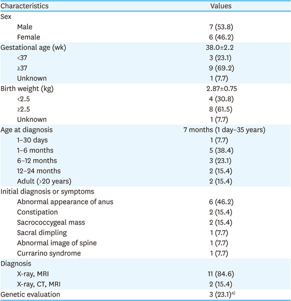
Table 1
Patient demographics (n=13)
All patients underwent a simple abdominal X-ray and magnetic resonance imaging (MRI) for the diagnosis of Currarino syndrome. Computed tomography (CT) was also done in two patients, who were diagnosed earlier within the study period, and they also had an MRI after CT.
Genetic evaluation was performed in three patients, but only one patient showed the MNX1 mutation. The associated anomalies were found in 11 patients, and the most commonly associated anomalies were urological anomalies.
The most common sacral anomaly was partial sacral agenesis (type III, 62%) followed by hemisacrum (type IV, 38%) (Table 2). No type I or II sacral anomalies were found. Meanwhile, the most common presacral mass was mature teratoma followed by anterior meningocele. No malignant features were observed in the pathological examination.
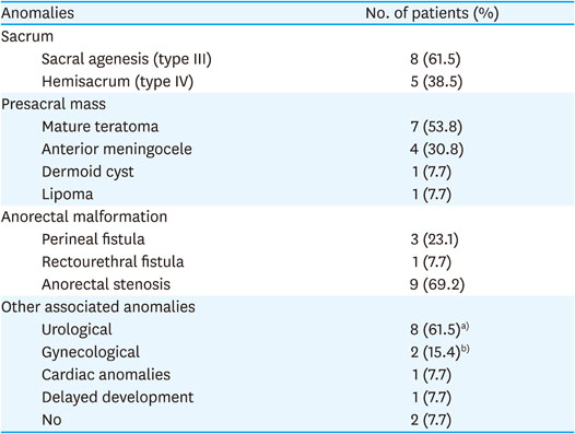
Table 2
Anomalies in Currarino syndrome (n=13)
In the ARM cases, most patients had anal stenosis (9/13), and all of the females in this study showed anorectal stenosis (6/6). Three other patients had low-type imperforate anus (perineal fistula), but one patient had high-type imperforate anus (rectoprostatic urethral fistula).
All of the cases underwent repair of ARM. Posterior sagittal anorectoplasty (PSARP) was the most common operative method; nine cases eventually underwent PSARP, but two of them underwent anoplasty and anal myectomy before undergoing PSARP, which in turn was performed as a re-do anoplasty. Two patients underwent a pull-through operation. In addition, four patients underwent colostomy before the operation for ARM during which the occurrence of Currarino syndrome was unknown.
Presacral mass excision was performed in 12 cases, except in one case. This case had presacral anterior lipomeningocele wherein neurological symptoms and lesion size were not indicated for surgery. In seven cases, presacral mass excision was combined with PSARP. Ten patients underwent neurosurgery involving detethering of the spinal cord with excision of meningocele, meningomyelocele, and/or film terminale lipoma.
We found various postoperative complications in seven patients; these complications include wound evisceration, anastomosis leakage after colostomy repair, cerebrospinal fluid (CSF) leakage after neurosurgery, recurrence of presacral teratoma, megarectum, and anal stenosis with iatrogenic rectovaginal fistula (Table 3).
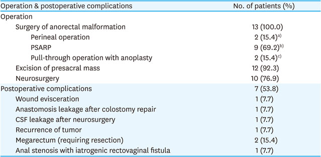
Table 3
Surgical treatment of Currarino syndrome (n=13)
One adult patient could not be followed up after the operation. The 12 other patients were followed up beyond the age of three and were evaluated for postoperative functional outcome. The median follow-up age was 8.1 years (range: 3.1–30.0 years).
Among the 12 patients, 4 patients did not show symptoms of functional abnormality, whereas 8 patients experienced constipation, fecal incontinence, urinary dysfunction, or gait disturbance. Seven of these eight patients had a defecation problem. Moreover, 3 patients had constipation, 5 had fecal incontinence, including 1 patient who had both symptoms. All constipation cases were classified as grade 1 or 2 and could be managed with laxatives. However, of the five patients with fecal incontinence, two had grade 3 soiling and ultimately underwent antegrade continence enema (ACE) procedures.
Another poor long-term outcome was urinary dysfunction as seen in five patients. Three of them underwent clean intermittent catheterization, and one patient required medication. In one case, Deflux® was injected due to vesicoureteral reflux. Four of them also had defecation problems, three of whom had gait disturbance and scoliosis requiring regular follow up (Table 4).
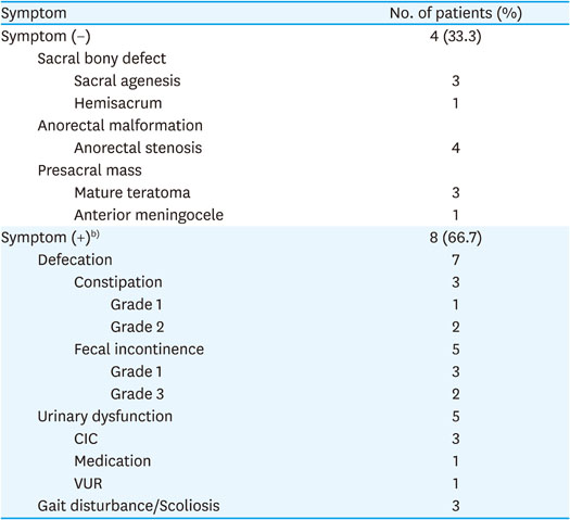
Table 4
Long-term functional outcome (n=12)a)
Since it was first described in 1981, Currarino syndrome became known to pediatric surgeons [1], and some studies described its characteristics and treatment outcomes [7, 10, 13, 14]. Nevertheless, it remains clinically unfamiliar that its diagnosis and treatment are still a challenge.
Our study showed some significant results in the diagnosis of Currarino syndrome. Currarino syndrome was diagnosed during the neonatal period in only one patient, whereas its diagnosis was delayed in the other patients. Currarino syndrome could be easily suspected because the characteristic appearance of sacrum (i.e., scimitar sacrum) can be observed in a plain abdominal film [9]. However, we overlooked this abnormal finding, indicating that a careful inspection of sacral anomalies in plain abdominal film is essential for the early diagnosis of this syndrome. The other reason for the delayed diagnosis was paying too much attention on the other anomalies associated with this syndrome. In this study, the most common initial symptom or diagnosis was ARM, which is characterized by the abnormal appearance of the anus, followed by constipation and sacrococcygeal mass. Consequently, seven patients underwent anoplasty, anal myectomy, or colostomy without the diagnosis of Currarino syndrome.
In this study, nine patients (69.2%) had anorectal stenosis, and this type of ARM was seen in all of the female patients. The incidence of anorectal stenosis is high in Currarino syndrome, and this finding is consistent with that reported by previous studies [10, 13, 14]. The reason as to why anorectal stenosis frequently occurs in Currarino syndrome is obscure, but this type of ARM is sometimes overlooked as a normal anus, leading to the delayed diagnosis of Currarino syndrome. Careful examination is thus required to avoid delayed diagnosis.
The purpose of the surgical treatment of Currarino syndrome is considerably straightforward, but we performed various surgical treatment sequences depending on the diagnostic sequence of Currarino syndrome. However, when possible, the co-operation of ARM and excision of presacral mass is a good treatment option. More than half of cases underwent PSARP and excision of presacral mass simultaneously, and no complication related to this co-operation occurred. However, we found various kinds of postoperative complications in seven patients, including wound evisceration, anastomosis leakage after colostomy repair, CSF leakage after neurosurgery, and recurrence of presacral teratoma, megarectum, and anal stenosis with iatrogenic rectovaginal fistula. We presume that the complex anomalies and rarity of this syndrome contributed to the high incidence of and to the different kinds of complications.
In this evaluation of postoperative functional outcomes, we found that most of the patients suffered from defecation difficulties and urinary dysfunction. Moreover, three patients had symptoms of gait disturbance and scoliosis. In defecation-related cases, fecal incontinence was more common than constipation, and two patients underwent ACE procedures. Although the functional outcome of Currarino syndrome has been reported [7, 10, 13, 15], there is a possibility that the long-term bowel and urinary dysfunction is underestimated. This study suggests that the postoperative clinical course is not usually favorable, and the patients will require care for their long-term functional dysfunction.
In this study, all patients had a preoperative MRI. Nowadays, MRI is one of the popular imaging techniques, and it is useful in characterizing and in determining the extent of lesions [16]. We believe that it should be performed preoperatively in all cases in conjunction with the use of plain abdominal film.
In this study, we could not discuss the genetic mutation of Currarino syndrome because only three patients underwent genetic evaluation, and only one of them showed the MNX1 mutation. Recently, the primary genetic cause of Currarino syndrome is known to be the pathogenic variants in MNX1 (previously known as HLXB9), and it was reported that 25%–30% of sporadic Currarino syndrome showed this type of mutation [3, 17]. We believe that the role of genetic mutation should be extensively evaluated in future studies.
This study has retrospective limitations, namely, the small number of patients and the heterogeneous surgical approach employed. However, we believe that our results, which were based on a relatively long-term follow up of functional outcome, will help manage Currarino syndrome.
In summary, the diagnosis of Currarino syndrome may be delayed when the sacral bony anomaly in a simple X-ray is not given attention. MRI evaluation is mandatory in patients who have ARM and sacral anomaly. The early diagnosis of this syndrome makes the co-operation of ARM and excision of presacral mass or another neurosurgery possible and can diminish unnecessary operation times. A careful follow up is essential because poor long-term functional outcomes are common.
Conflict of Interest:No potential conflict of interest relevant to this article was reported.
Author Contributions:
Conceptualization: K.H.J., O.J.T.
Data curation: K.H.J., H.I.G., I.K., H.S.J., O.J.T.
Methodology: K.H.J.
Supervision: O.J.T.
Validation: H.I.G., I.K., H.S.J.
Writing - original draft: K.H.J.
Writing - review & editing: O.J.T.
