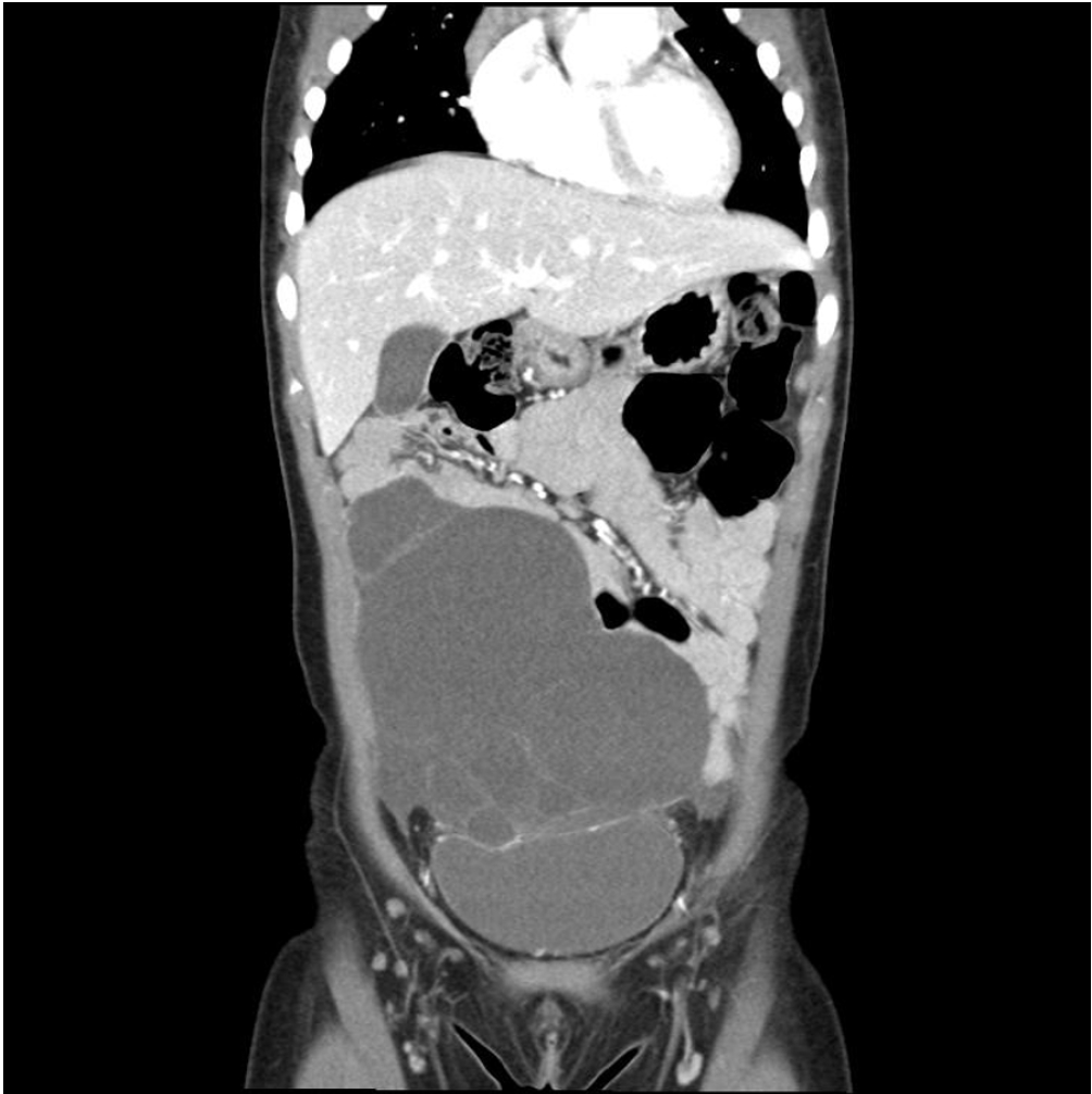
Benign cystic mesothelioma (BCM) is a rare intra-abdominal tumor and is particularly uncommon in pediatric patients. Its nonspecific clinical and radiological features often make preoperative diagnosis challenging. We report the case of a 4-year-old girl who presented with acute abdominal pain and vomiting. Computed tomography revealed a large, multiloculated cystic mass occupying the lower abdomen, which was initially suspected to be a lymphatic malformation. During laparoscopic exploration, a hemorrhagic, multiloculated cystic mass with hemoperitoneum was identified and completely resected with preservation of the right ovary. Histopathological examination confirmed BCM, showing predominant clusters of epithelioid cells interspersed with cyst-like spaces on hematoxylin and eosin staining. The patient recovered uneventfully and had no recurrence during 30 months of follow-up. This case describes an unusual presentation of BCM as an acute abdomen complicated by hemoperitoneum in a child and emphasizes the importance of surgical exploration and histopathological evaluation in establishing the diagnosis.
Purpose The International Society for the Study of Vascular Anomalies (ISSVA) classification is crucial in diagnosing vascular anomalies (VAs), surpassing the International Classification of Diseases 10th Revision. This study aims to reevaluate diagnoses using ISSVA criteria and explore diagnostic patterns.
Methods Analyzing 138 pediatric VA patients diagnosed via magnetic resonance imaging from 2018 to 2023 at Asan Medical Center, we reviewed clinical, imaging, pathology, and genetic data. Diagnoses were revised per 2018 ISSVA criteria, assessing discrepancies.
Results Among 133 VA cases, 125 were malformations and eight were tumors, mostly in the head and neck. Clinical and imaging diagnoses disagreed in 51 cases. Some initially complex malformations were simplified. Lymphatic malformation cases shifted to VAs and vascular tumors were identified post-initial diagnosis.
Conclusion Accurate diagnosis of VAs is essential for prognosis, treatment planning, and predicting outcomes. However, 14.2% of patients showed discordance between clinical diagnoses and imaging findings. Capillary malformations were often overlooked in imaging but became evident with relevant clinical findings. Adopting a multidisciplinary approach and a unified diagnosis based on ISSVA classification is crucial for clearly defining VAs.
Since the first introduction of robotic surgery systems in Korea in 2005, there has been a gradual increase in the number of robotic surgeries performed. However, robotic liver resection is one of the most complex procedures, and its application, especially to children, is still limited. Therefore, in this study, we aim to present our experiences with 2 pediatric patients who underwent robotic liver resection in Asan Medical Center and discuss the safety and feasibility of robot-assisted hepatectomy in pediatrics.
Purpose Complicated vascular anomalies, characterized by encasing vital organ or diffusely locating unresectable lesion, pose therapeutic challenges with limited response to conventional treatment such as surgical resection or sclerotherapy. Sirolimus, an mammalian target of rapamycin inhibitor, has shown promising therapeutic effects in patients with vascular anomalies by inhibiting vascular endothelial growth factor, as reported in several studies. Here, we analyzed the treatment outcomes of patients who received sirolimus for complicated vascular anomalies at our institution.
Methods Patients treated with sirolimus at the Department of Pediatric Surgery, Asan Medical Center from January 2018 to December 2021 were included. Sirolimus was administered twice daily at a dose of 0.8 mg per body surface area (BSA), with dose adjustments to achieve a target drug concentration of 8–12 ng/mL. Adverse drug effects and therapeutic responses were periodically assessed. Treatment efficacy was evaluated based on clinical findings pre- and post-sirolimus administration, absolute volume reduction of lesions through imaging tests (magnetic resonance imaging; MRI), and relative volume reduction adjusted to the patient's BSA.
Results There were 16 females (50.0%) and 16 males (50.0%), with a median follow-up period of 41 months after sirolimus administration. Vascular anomaly types included lymphatic malformations (41%), venous malformations (28%), lymphovenous malformations (19%), and others (12.5%). The most common adverse effect was oral ulcer (6 patients). MRI volumetry revealed volume decreases in 17 patients (53.1%) with 22 patients (71%) exhibited lesion decreases relative to BSA. Notably, 9 patients (28.1%) had markedly decreased volume reduction based on absolute volume, and 12 (38.7%) based on volume compared to BSA.
Conclusion Over a 2-year follow-up, sirolimus was effective in treating patients with complicated vascular anomalies, when administered with cautious consideration of side effects. A multidisciplinary approach is needed for evaluating treatment outcomes in these patients, necessitating further long-term research on adverse effects.

 First
First Prev
Prev

