

Purpose
Report of a nationwide survey on lymphangioma conducted by the Korean Association of Pediatric Surgeons (KAPS) in 2019.
Methods
The authors reviewed and analyzed the clinical data of pediatric patients who started treatment for lymphangioma in hospitals of KAPS members from 2011 to 2013. Their follow-up data is also included in the study.
Results
A total of 532 patients with lymphangioma from 18 institutes were registered for the study. The results were discussed at the 35th annual meeting of KAPS, which was held in Gyeongju on June 13–14, 2019.
Conclusion
This study provides general information on lymphangioma and comprehensive treatment outcomes for this disease. The study is expected to be an important reference for improving pediatric surgeons’ understanding and treatment of lymphangioma.
Report of a nationwide survey on lymphangioma conducted by the Korean Association of Pediatric Surgeons (KAPS) in 2019.
The authors reviewed and analyzed the clinical data of pediatric patients who started treatment for lymphangioma in hospitals of KAPS members from 2011 to 2013. Their follow-up data is also included in the study.
A total of 532 patients with lymphangioma from 18 institutes were registered for the study. The results were discussed at the 35th annual meeting of KAPS, which was held in Gyeongju on June 13–14, 2019.
This study provides general information on lymphangioma and comprehensive treatment outcomes for this disease. The study is expected to be an important reference for improving pediatric surgeons’ understanding and treatment of lymphangioma.
Lymphangioma or lymphatic malformation includes a wide spectrum of abnormalities because it can be found in any region of the body and has a variety of appearances [1, 2, 3, 4]. Although pediatric surgeons occasionally encounter this disease, the incidence of lymphangioma is relatively low. This means that it takes a long time for surgeons to accumulate experience in the treatment of lymphangioma. Therefore, a nationwide study is helpful for surgeons to understand this disease and choose optimal treatment methods. The 2019 annual nationwide survey of the Korean Association of Pediatric Surgeons (KAPS) was on the subject of lymphangioma, and the results were first discussed at the annual meeting of KAPS in Gyeongju on June 13–14, 2019.
The authors reviewed and analyzed the clinical data of patients who started treatment for lymphangioma in KAPS members’ hospitals from 2011 to 2013. Their follow-up data were also included in the study. We used Microsoft Access 2016® (Microsoft, Redmond, WA, USA) for patient registry and data collection. All the data were analyzed using the IBM SPSS version 23 statistical software package (IBM Co., Armonk, NY, USA).
A total of 532 patients with lymphangioma from 18 institutes were registered for the study (Fig. 1). Although 550 patients were reported, 18 patients were reported by 2 institutes. Therefore, we unified the data of these patients. The patients’ demographics and clinical characteristics are summarized in Tables 1 and 2. Age at diagnosis varied; 39.3% of patients (209/532) were diagnosed in the infant period, and 25% (133/532) were diagnosed after the age of 5. Only 12.8% of patients (68/532) were diagnosed postnatally. The majority of patients (81.0%) were treated by pediatric surgeons. Most patients (94.9%) had a lesion at a single site, but 27 patients had multiple lesions at more than one site. The most common symptom was mass, followed by pain. The most common imaging study was ultrasound, followed by magnetic resonance imaging. We classified lymphangioma with a diameter of more than 1 cm as macrocystic. Macrocystic lymphangioma was the most common type of lymphangioma, and the median diameter size was 4.8 cm.
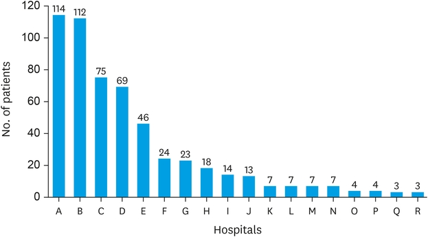
Fig. 1
Hospital distribution of patients with lymphangioma.
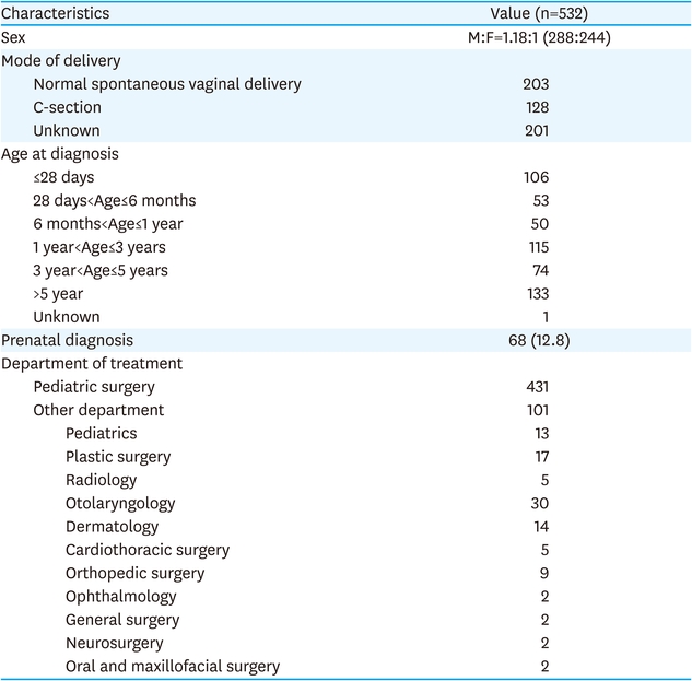
Table 1
Patient demographics
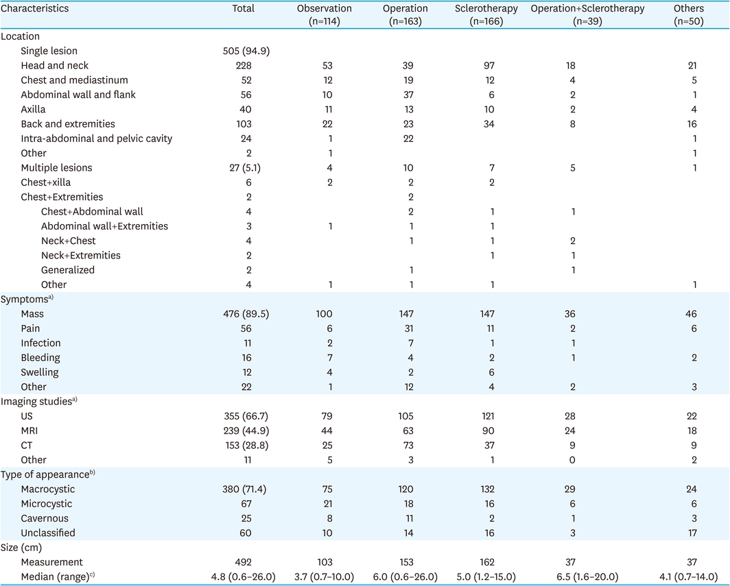
Table 2
Clinical characteristics
We grouped treatment methods into 5 categories: observation only; operation; sclerotherapy; operation and sclerotherapy; and other treatments (Fig. 2). Operation with observation and sclerotherapy with observation were classified as operation and sclerotherapy, respectively.
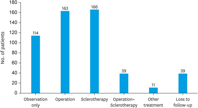
Fig. 2
Treatment methods.
Patients who underwent observation only are summarized in Table 3. A total of 114 patients (21.4%) did not undergo any specific treatment, and the median observation period was 316 days. Among the patients in this group, 67.9% (88/114) showed grossly normal or partially improved appearance.
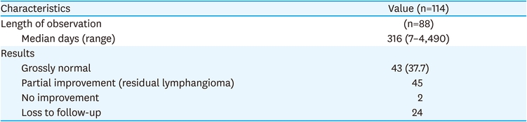
Table 3
Observation only
A total of 163 patients (30.6%) underwent an operation. The predominant operation site was the head and neck, followed by the abdominal wall and flank. Characteristically, lesions in the intra-abdominal and pelvic cavity were more common in this group compared to the other treatment method groups. The most common histopathological diagnosis was lymphangioma, followed by hemangiolymphangioma. Most patients underwent complete excision in a single operation. Only 8% (13/163) of patients required more than 1 operation. Three patients underwent sclerotherapy during operation. Postoperative complications were not common; only 7.4% of patients experienced complications, and complications were of various kinds. After operation, 84.5% (138/163) of patients showed grossly normal results or no residual lesions (Table 4).
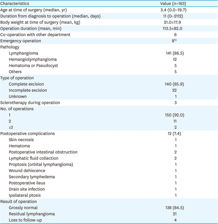
Table 4
Operationa)
A total of 166 patients (31.2%) underwent sclerotherapy, which is similar to the number of patients who underwent operation. The most common sclerosing agent was OK-432, followed by bleomycin and doxycycline. General anesthesia was required in the majority of patients, and more than one-third of patients required multiple sessions of sclerotherapy. Complications after sclerotherapy were extremely rare; only 3 patients experienced complications. After sclerotherapy, 57.2% (95/166) of patients showed grossly normal results (Table 5).
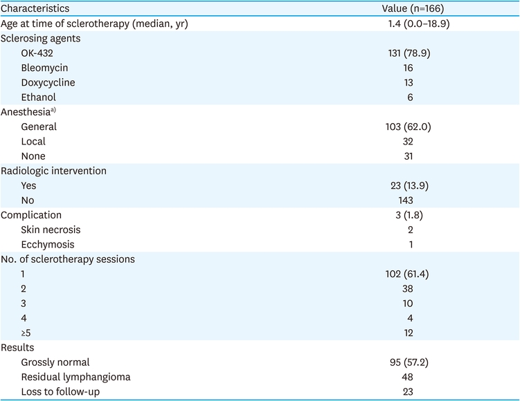
Table 5
Sclerotherapy
Thirty-nine patients (7.3%) underwent operation and sclerotherapy separately, not simultaneously. The majority of these patients underwent sclerotherapy either before or after operation, but 3 patients underwent sclerotherapy both before and after operation. Multiple sclerotherapy sessions were required in 51.3% (20/39) of patients. After treatment, 53.8% (21/39) showed grossly normal results (Table 6).
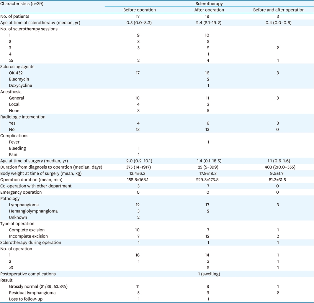
Table 6
Operation and sclerotherapy
A total of 11 patients underwent other treatments. Of these, 4 had drug treatment (2 cases of steroid medication and 2 cases of antibiotic medication), 4 underwent ultrasound-guided aspiration, and 3 had CO2 laser abrasions. Only 2 patients showed grossly normal results at follow-up.
Although lymphatic malformations can occur in any anatomic region, most clinical studies of lymphangioma, or lymphatic malformation, focus on lymphatic-rich areas, such as the head and neck, the axilla, and the mediastinum [3, 5, 6]. However, understanding the overall clinical characteristics of lymphangioma is important because the basic concepts in the treatment of lymphangioma are similar, regardless of the lesion site [2, 4]. The significance of this study lies not only in the fact that it is the first nationwide survey on lymphangioma in Korea but also in the fact that it integrates clinical characteristics and treatment results of patients with lymphangioma.
The results of this study are consistent with those of previous studies with respect to the most common locations of lesions, the major symptoms, the kinds of imaging studies, the types of appearances [1, 7], and favoring of OK-432 as a sclerosing agent [8, 9, 10]. However, in contrast with other studies, we included observation without other treatments as a method of treatment [6]. We believe that the benign characteristic of lymphangioma and the possibility of spontaneous regression makes observation a viable treatment option [11].
Among the treatment methods considered, the best results were obtained from operation, followed by sclerotherapy, co-treatment with operation and sclerotherapy, and observation. However, the correlation between the choice of treatment method and the difficulty of treatment should be considered when interpreting these results. The difficulty of treating lymphangioma generally varies significantly depending on whether it takes a simple or complex form [5, 12]. We think it was possible that we avoid the operation in complex form of lymphangioma, and non-operative treatment methods were selected as the preferred treatment method over operation. In this study, we did not distinguish between simple and complex forms of lymphangioma, which is one of the limitations of this study.
Other limitations are that we did not include long-term follow-up results, and we did not compare results according to the area where the lesion occurred. These issues should be addressed in future studies. Despite its limitations, this study provides general information on lymphangioma and comprehensive treatment outcomes. Therefore, we expect it will be an important reference for pediatric surgeons seeking to better understand lymphangioma and treatment options for this disease and that it will ultimately lead to better outcomes.
Conflict of Interest:No potential conflicts of interest relevant to this article are reported.
